Case Report: Removal of Textured Implants After 10 Years With Capsular Contracture
MD, MSc. Ho Cao Vu
Practising certificate No. 028475/BYT-CCHN, issued by the Ministry of Health on 28 June 2018. Specialist in general surgery and plastic–aesthetic surgery, with more than 15 years in plastic and aesthetic surgery and more than 20 years of general surgical practice covering benign breast disease, breast cancer, and abdominal wall and abdominal surgery at Cho Ray Hospital. In 2010 he trained in post-cancer breast reconstruction at MD Anderson Cancer Center, Houston, Texas, USA, under the direct supervision of Prof. David Chang. He currently practises in the Department of Surgery, Nam Sai Gon International General Hospital, where he personally performs plastic and aesthetic procedures. By 2025 he reported more than 1,000 breast implant removal cases and 3,000 plastic and aesthetic procedures, using the ultrasonic surgical scalpel.
Updated September 2026. Case details are published with the patient’s consent and with identifying information removed. Clinical context cross-checked against US Food and Drug Administration (FDA) information on breast implant risks and imaging, and American Society of Plastic Surgeons (ASPS) patient material.
Short answer: a woman in her forties asked for removal of textured 300 cc implants placed 10 years earlier, because of heaviness, breast ptosis and a tightening sensation on the right. Pre-operative MRI showed a folded implant with the shell apparently intact. At surgery the capsule measured 2–3 mm thick on both sides, the pocket was narrow, and the implant was found to have ruptured inside its capsule, with yellow fluid at the base of the pocket. Both implants and both capsules were removed, capsule tissue was sent for histology, and no lift was performed at the same operation — a decision made deliberately, and explained below.
Why this case is worth publishing
Three findings in this case recur often enough in explant practice to be worth setting out in full.
The first is the gap between imaging and operative findings: the MRI report described a fold with an apparently intact shell, and the rupture was identified only once the pocket was opened. The second is the thickness and distribution of the capsule — thickest on the posterior wall, where a narrow pocket had been pressing the implant against the chest wall for a decade. The third is the decision not to perform a lift at the same operation, which the patient specifically asked about.
This report follows the case through assessment, decision-making, operative findings and the reasoning behind staging. For the general framework of explant surgery, see our complete guide to breast implant removal.
Presentation
| Detail | |
|---|---|
| Time since augmentation | 10 years |
| Implants | Textured surface, 300 cc each side |
| Main complaints | Heaviness, breast ptosis, tightening sensation on the right |
| Original indication | Augmentation for breast ptosis noted before the first operation |
| Reported at first consultation | Patient recalled feeling at the time that the recommended volume was large for her frame, but did not raise it |
| Pre-operative imaging | MRI: implant fold; shell reported as apparently intact |
| Examination | Right breast firmer than left, with changes consistent with contracture |
The patient had lived with the implants for a decade without any earlier assessment. That is a common pattern: symptoms develop gradually, and the change is attributed to age or weight rather than to the device.
Assessment
Step 1 — History. Date of the original operation, implant type and surface, volume, and the reason augmentation was chosen. Here the original indication was breast ptosis treated with volume alone, which is relevant to what the breast would look like once the volume was removed.
Step 2 — Examination. Both breasts were assessed for firmness, shape, implant position and symmetry. The right breast was firmer, with a tightening sensation the patient described as uncomfortable rather than painful.
Step 3 — Imaging. Dedicated breast MRI was requested rather than ultrasound or mammography alone. The FDA advises periodic imaging for people with silicone implants even without symptoms, as set out in its risks and complications information; our article on breast implant screening for silent rupture explains what each modality can and cannot show. The MRI described a fold in the implant, and the shell was reported as apparently intact.
Step 4 — Explaining what a fold means over time. A folded implant creates a crease that is repeatedly stressed. Over years, folds are associated with progression to higher-grade contracture and with shell failure at the crease. This was the basis for recommending removal rather than continued observation.
Step 5 — Agreeing the plan, including what would not be done. Removal of both implants with capsule removal, capsule sent for histology, and no simultaneous mastopexy. The reasoning for staging is set out below, and was discussed before the operation so the appearance immediately afterwards would not come as a surprise.
The decision not to lift at the same operation
The patient asked whether a lift could be performed during the same operation. The answer in this case was that it should not be, for reasons specific to her findings.
The breast tissue had been stretched under sustained tension for 10 years, so the soft tissues were not in a stable state to plan a lift against. The capsule had to be removed completely and sent for histology, which by itself adds operative time to an already substantial dissection. Combining a full capsule removal with a mastopexy would have lengthened the anaesthetic and been planned on tissues whose final position was not yet known.
The plan was therefore to remove the implants and capsules, allow roughly 6 months for the tissues to settle, and then assess whether any contouring procedure was wanted at all. Some patients decide at that point that they no longer want further surgery. The trade-offs between removal alone, removal with a lift and removal with replacement are set out in our complete guide; the general point is that a breast which had ptosis before augmentation is likely to show it again once the volume is taken out, and the patient was shown what that would look like beforehand.
Operative findings
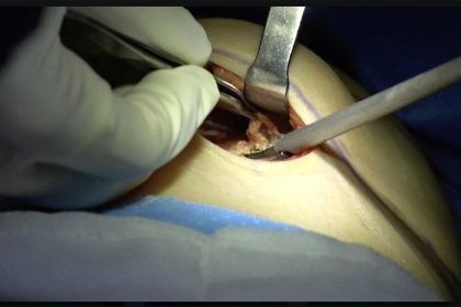
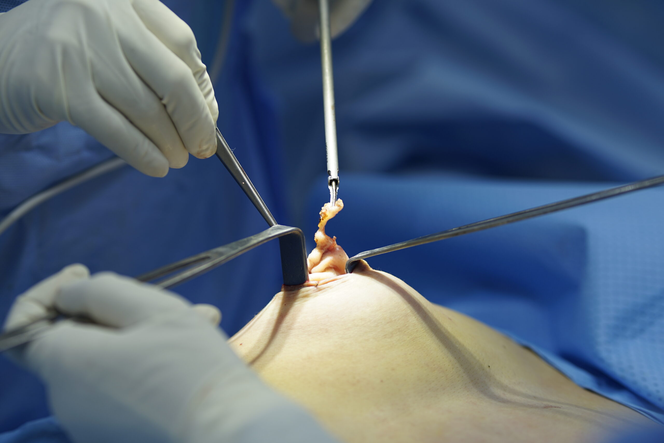
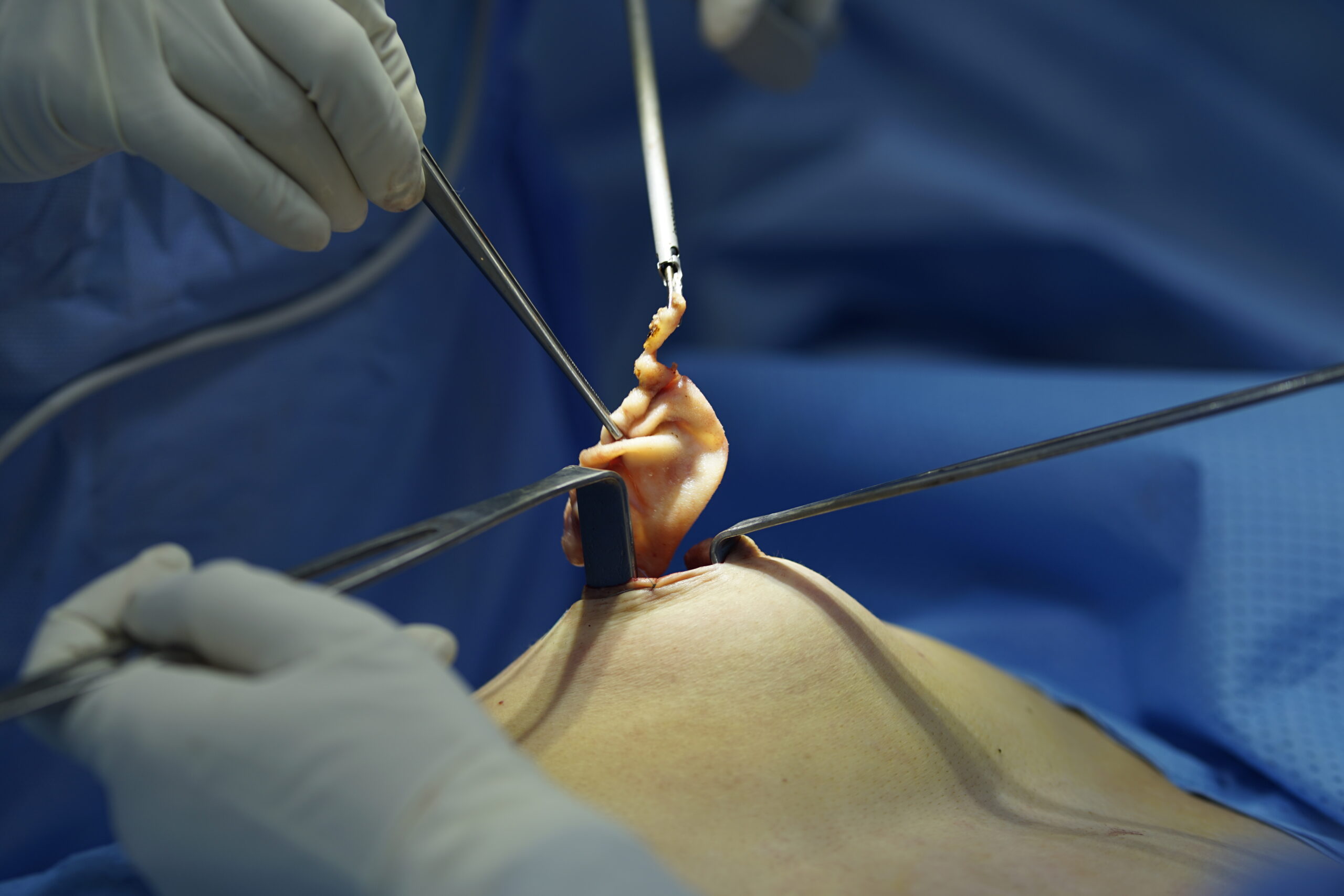
The operation was performed under general anaesthesia with endotracheal intubation, patient supine. A periareolar incision was used on both sides. Dissection into the partial subpectoral pocket was carried out with the ultrasonic surgical scalpel, which divides and coagulates tissue in the same pass.
Findings recorded at surgery:
- The pocket was narrow relative to the implant that had been placed in it.
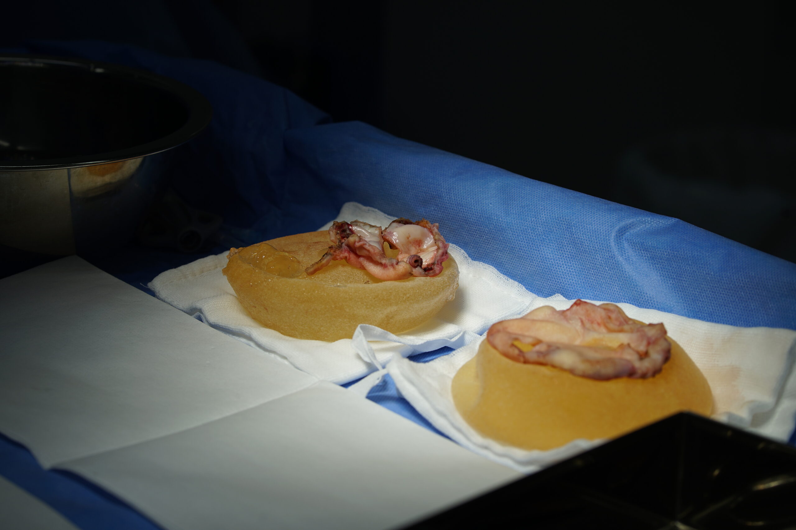
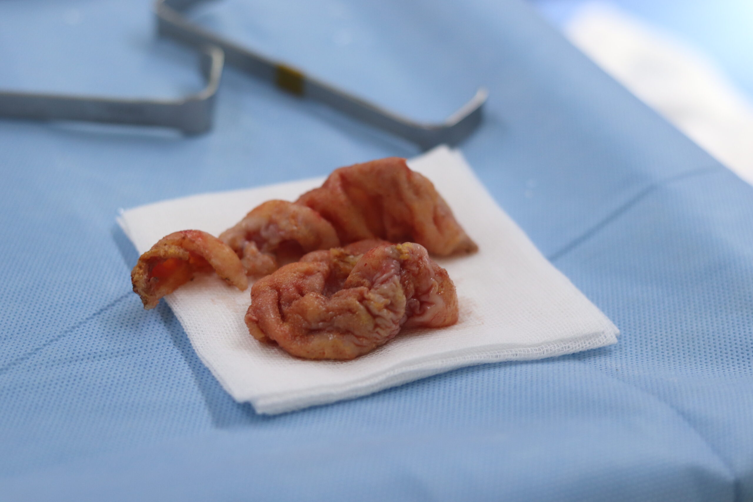
- The posterior capsule was markedly thickened and rigid — thicker than the anterior capsule, and the reason the base of the implant appeared smaller than its outer rim.
- Capsule thickness measured 2–3 mm on both sides.
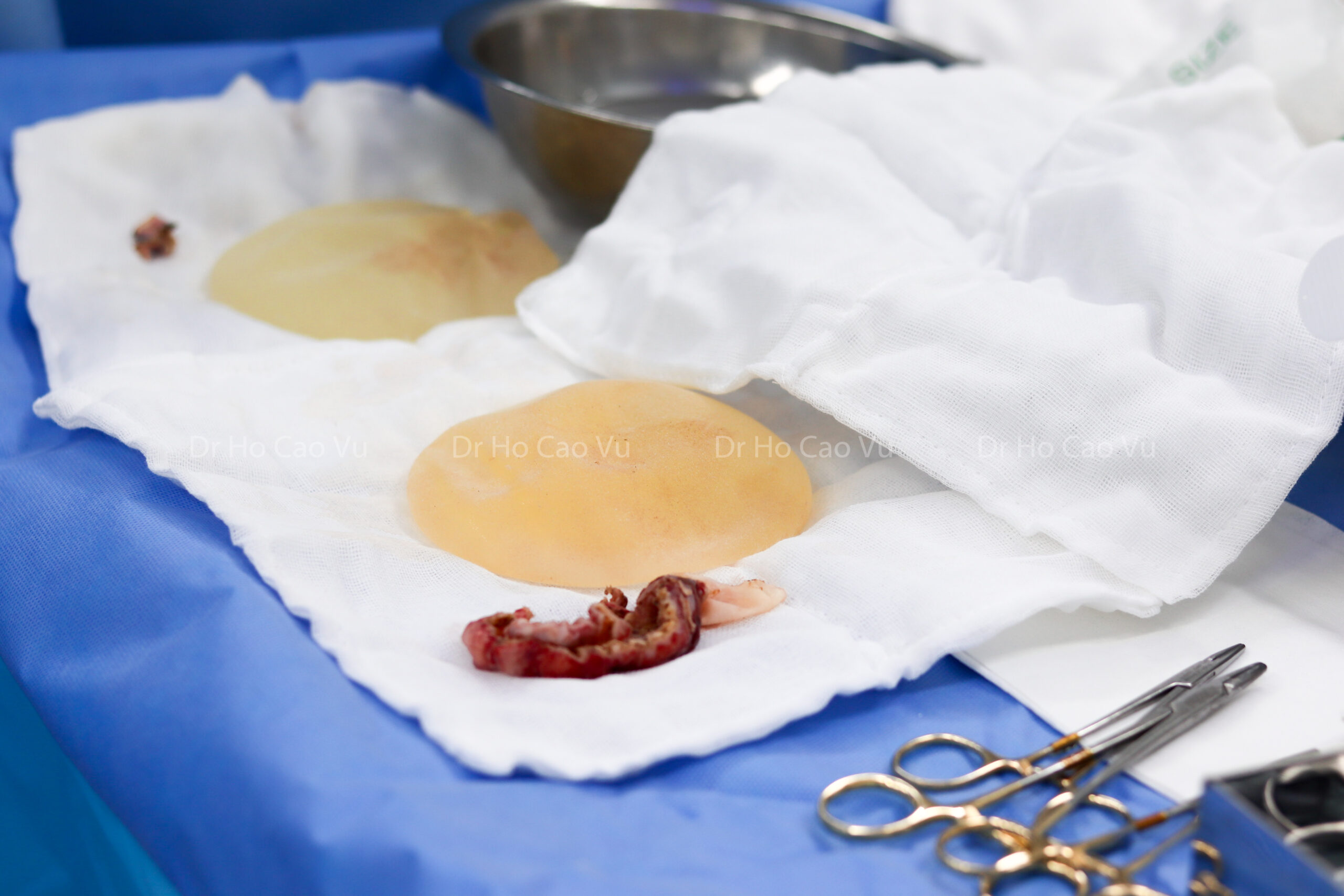
- The implant was folded, with a pocket of tissue lying between the two folds.
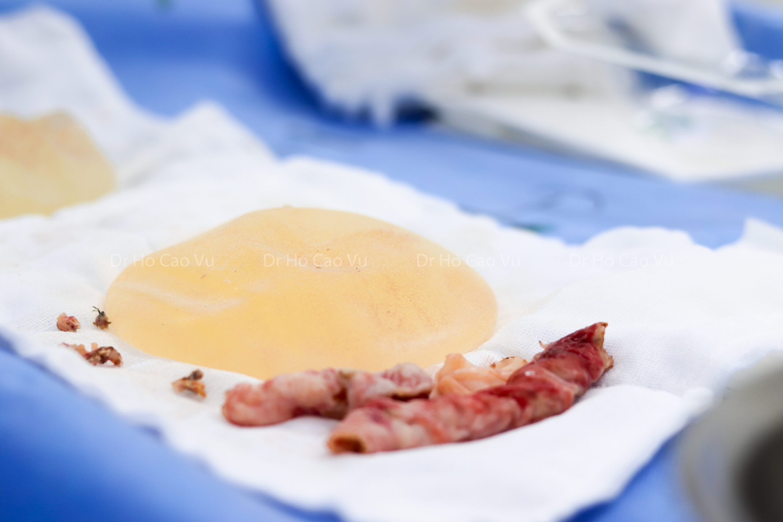
- The implant had ruptured inside the capsule, and yellow fluid was present at the base of the pocket — neither finding predicted by the pre-operative MRI report.
Both implants were removed. The capsules were excised on both sides and sent for histopathology. The wounds were closed in layers.
What the findings mean
Imaging is a guide, not a verdict. MRI is the most sensitive available test for silicone implant rupture, and here it identified the fold correctly. It did not identify that the shell had already failed within the capsule. This is why the operative plan included capsule removal and pathology regardless of what the imaging suggested, and why a contingency was discussed in advance. The way silent rupture presents is covered in our article on silent breast implant rupture.
Pocket size and capsule thickness are related. The thickest capsule lay where the implant had been pressed against the chest wall in a pocket that was narrow for its volume. The patient’s own recollection — that the recommended volume seemed large for her frame — matches what was found a decade later.
Fluid in the pocket is assessed, not ignored. Fluid found around an implant, particularly years after the original surgery, is documented and the capsule is sent for histology. Sending capsule tissue for pathology is routine practice in explant surgery, and it is the route by which uncommon implant-associated conditions are identified.
How the capsule is graded, and where this case sat
Capsular contracture is graded clinically on a four-point scale, and the grade is one of the inputs into whether removal is recommended:
| Grade | What is found |
|---|---|
| I | Breast is soft; shape and size appear natural; no effect on the result of the augmentation |
| II | Slight firmness, with the shape still normal |
| III | Moderate firmness with visible change in shape |
| IV | Firm, distorted breast, often with pain |
The right breast in this case was firmer than the left, with the tightening sensation the patient described, and the operative findings — a rigid posterior capsule measuring 2–3 mm — matched a capsule that had been thickening for a long period. Grading is covered in more detail in our article on capsular contracture after breast augmentation.
Two mechanisms that recur in cases like this
A capsule forms around every implant within roughly 4–6 weeks of surgery; that is a normal healing response, and in most women the capsule stays thin and is not felt. Two situations recur in cases where it does not, both of them visible in the findings here.
The first is a pocket that is narrow relative to the implant volume. When the space created at the original operation is smaller than the device it has to hold, the implant is under continuous pressure against the chest wall. Over years this is associated with a thicker posterior capsule — which is precisely where the capsule was thickest in this case, and consistent with the patient’s own recollection that the recommended volume seemed large for her frame.
The second is tissue handling during the original pocket dissection. A rough dissection is associated with more post-operative pain, more bleeding and fluid production, slower healing, and late complications including contracture. This is one of the reasons the ultrasonic scalpel is used at this practice for both augmentation and removal: it divides and coagulates in the same pass, which limits the number of separate steps needed to control small bleeding points across a large dissection.
Neither mechanism can be confirmed retrospectively in an individual patient a decade later. They are set out here because they inform how the next operation is planned — in this case, why the surgeon was unwilling to combine a full capsule removal with a lift in one sitting.
Recovery and staging
The patient was managed as a standard explant recovery: supportive garment, activity restricted in the early period and increased in stages, and review appointments to monitor healing. Return to desk-based work is generally discussed in the range of 1–2 weeks and a return to full exercise over 4–6 weeks, individually assessed — these are planning figures used at consultation, not fixed promises, and they vary with the extent of the dissection and individual healing. The ASPS patient information on breast implant removal sets out the general recovery framework.
The staged plan means the question of contouring is revisited at around 6 months, when the tissues have settled and the result of removal alone can be seen. A comparable staged decision in a breast with grade IV contracture is described in our case report on removal of Polytech textured implants after 11 years.
Points other patients ask about after reading a case like this
- “My implants feel fine — should I have imaging anyway?” The FDA recommends periodic imaging for silicone implants in the absence of symptoms, because rupture within the capsule can develop without a change the patient can feel.
- “Does a thicker capsule mean something was done wrong?” Capsule thickness reflects several factors, including pocket dimensions relative to implant volume, the healing response and time in place. It is one finding among several, interpreted together rather than alone.
- “If my implant is folded, does it have to come out?” Not in every case. A fold is one factor weighed together with symptoms, contracture grade and how long the implant has been in place. Our comparison of surgical and non-surgical options for capsular contracture sets out how that judgement is made.
Frequently asked questions
Was this rupture visible on MRI before surgery? No. The MRI report described a fold with an apparently intact shell; the rupture inside the capsule was identified during the operation. Imaging guides planning, and the operative plan allowed for findings to differ.
Why was the capsule sent to pathology? Sending capsule tissue for histology is routine in explant surgery. It documents what was removed and is the route by which uncommon implant-associated conditions are identified, particularly where there is fluid in the pocket.
Why were the implants not replaced? The patient’s goal was removal. Where a patient wants to keep volume, replacement is discussed as a separate option with its own assessment; that was not the request here.
Will the breast look the same as before the original augmentation? Not exactly. Skin and breast tissue change over 10 years under sustained volume, and the breast had ptosis before the first operation. The appearance after removal was discussed with photographs at consultation so expectations were set before surgery, and the option of a staged procedure remains open.
How long is the wait before a second-stage procedure? In this case, about 6 months. The interval allows swelling to resolve and tissues to settle so that any second procedure is planned against a stable starting point.
Is textured surface relevant to the decision? Implant surface is recorded because it is relevant to surveillance and to the rare implant-associated conditions described by regulators. In this case the reason for removal was the combination of symptoms, fold and contracture, and the operative decisions followed those findings.
Summary
A textured 300 cc implant in place for 10 years, in a pocket that was narrow for its volume, produced a thickened capsule of 2–3 mm, a fold, and a rupture that imaging had not identified. Both implants and capsules were removed, capsule tissue went for histology, and a lift was deliberately staged rather than combined — because the tissues had been under tension for a decade and their final position was not yet known. The points that carry over to other patients are that imaging guides but does not settle the operative plan, that capsule findings tend to reflect the relationship between pocket and implant volume, and that staging is sometimes the way to get a better result rather than a delay.
References
- Risks and Complications of Breast Implants — U.S. Food and Drug Administration
- Breast Implant Surgery — U.S. Food and Drug Administration
- Breast Implant Removal — American Society of Plastic Surgeons
- Questions and Answers about Breast Implant-Associated Anaplastic Large Cell Lymphoma (BIA-ALCL) — U.S. Food and Drug Administration