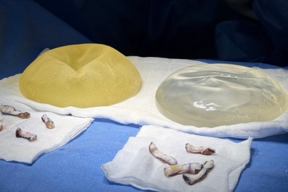
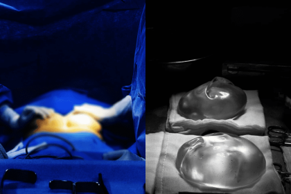
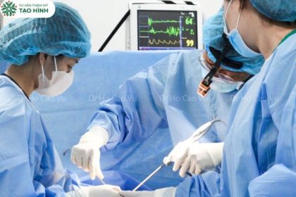
Silent Infection Found During a Third Breast Augmentation: What the Pocket Revealed
Short answer: a breast implant pocket can look normal on ultrasound and return a negative infection test while still containing active inflammation. In the case below, a patient cleared for her third augmentation was found — only once the pocket was opened with an ultrasonic scalpel — to have clumped white pseudomembrane on the left and yellow discharge on the right. The planned implant placement was abandoned mid-operation, the family was telephoned, and the operation became a washout with drainage and culture instead.
Last reviewed September 2026. Medically reviewed by MSc, MD Ho Cao Vu — Practising Certificate No. 028475/BYT-CCHN. More than 15 years in aesthetic plastic surgery and more than 20 years of general surgical experience at Cho Ray Hospital; trained in post-cancer breast reconstruction at MD Anderson Cancer Center, USA.
Why a negative test is not the same as a clean pocket
Patients preparing for revision breast surgery are often reassured by two things: a breast ultrasound that reports no collection, and a blood test that shows no infection. Both are useful. Neither is conclusive.
The FDA lists infection, capsular contracture and implant rupture among the recognised risks of breast implants, and notes that some complications are found only at revision surgery. Dr Ho Cao Vu notes that low-grade, walled-off inflammation inside an old capsule can persist without producing the systemic signals a blood test detects, and without forming a collection large enough for ultrasound to flag. The material sits in the recesses of the pocket — thin, adherent, easy to miss — and it only becomes visible when the pocket is opened.
This matters because placing a new implant into such a pocket is what turns a manageable problem into a repeating one. The article below follows a real revision case in which that decision was made correctly, in theatre, against the original surgical plan.
The case: Ms TT, born 1990
A three-operation history
Ms TT had a long and difficult implant history before she reached Dr Vu:
- April 2022 — first augmentation at another facility. She developed an abscess and the implants had to be removed.
- December 2023 — second augmentation. She developed a seroma. The surgeon removed the implants, placed a drain and sent her home.
- Recently — she wished to try again. A breast ultrasound was performed and an infection test returned negative.
On the strength of those results she was scheduled for implant placement on 17 December 2024, with Dr Vu operating personally.
What the surgeon found
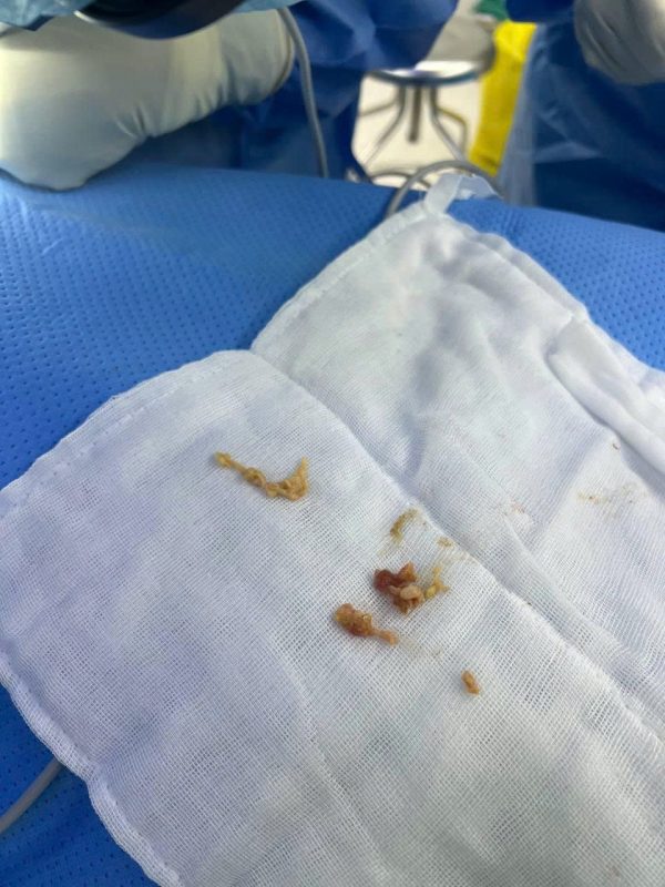
After dissecting into the pocket with the ultrasonic scalpel, Dr Vu found abnormalities that neither the ultrasound nor the blood test had shown.
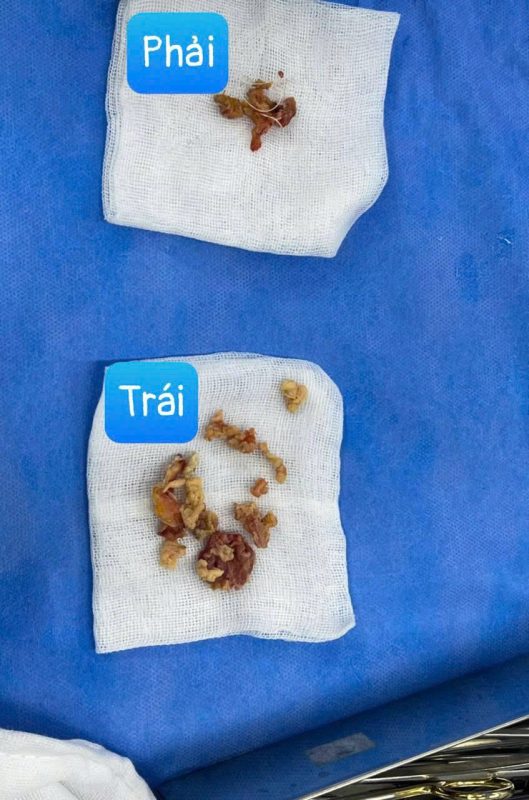
A pocket had formed earlier, with a thin shell whose surface bore scattered pseudomembrane in the recesses of the pocket. Specifically:
- Left breast pocket: a great deal of clumped white pseudomembrane.
- Right breast pocket: yellow discharge together with clumped pseudomembrane.
This was the result of inadequate cleaning of the pocket at a previous operation, which had caused intracapsular inflammation.
There was a second finding. On the left, the entire lower part of the pectoralis major over ribs 4, 5 and 6 was still intact and had not been divided. In a submuscular augmentation, failure to release the lower muscle attachments constrains the implant and prevents it sitting correctly in the lower pole — a structural problem sitting alongside an inflammatory one.
The decision made in theatre
The original plan was implant placement. Dr Vu abandoned it.
He telephoned the family, explained the findings and obtained agreement to change the operation. The revised procedure was:
- Break down the recesses of the pocket where pseudomembrane had collected.
- Remove part of the pocket in the lower portion on both the right and left sides.
- Irrigate with hydrogen peroxide and Betadine, then wash out again thoroughly with saline.
- Place a post-operative irrigation drain.
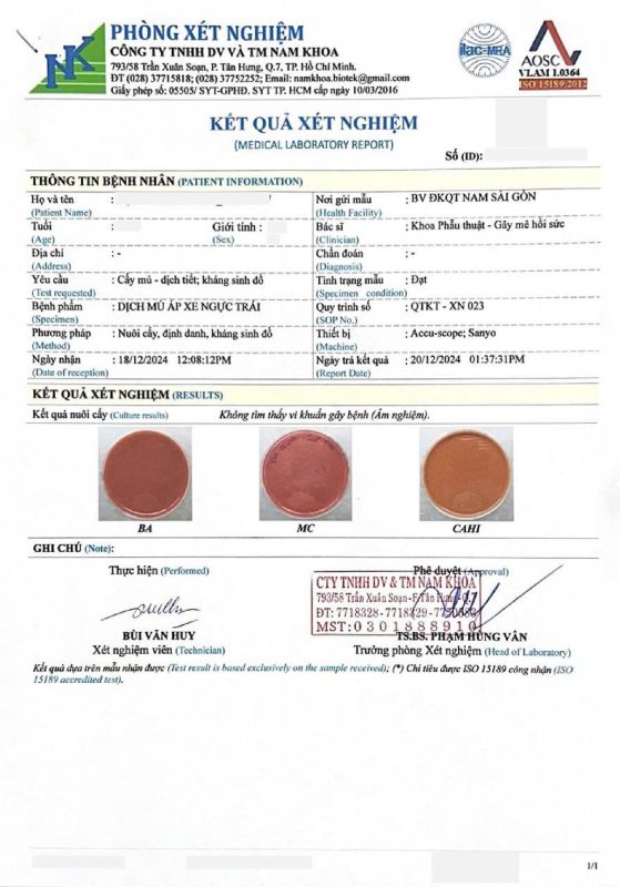
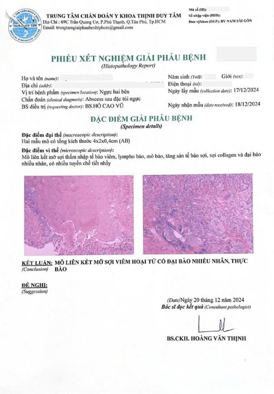
- Send the pseudomembrane for antibiotic sensitivity culture, histopathology, and testing for tuberculosis.
No implant was placed.
Why the operation was changed rather than completed
Placing an implant into a pocket containing pseudomembrane and discharge would have been the shorter path on the day and the longer path afterwards.
Inflamed tissue does not form a stable pocket. An implant needs a capsule that adheres and settles predictably. A pocket with retained inflammatory material continues to produce fluid, which is how the December 2023 seroma is likely to have arisen.
A foreign body in an inflamed field concentrates the problem. As covered in the discussion of biofilm in implant infection, bacteria adhering to a device surface become substantially harder to eradicate than the same organisms in tissue alone.
The pattern was already established. Two previous operations at other facilities had ended in abscess and seroma respectively. Repeating the same step a third time, into a pocket now visibly abnormal, would most likely have produced the same result.
Dr Ho Cao Vu notes that the most difficult conversation in revision surgery is the one that happens mid-operation, when the plan the patient consented to is no longer the right plan. Making that call is what protects the possibility of a good result later.
Why the specimen was sent for three separate tests
Sending pseudomembrane for antibiotic sensitivity culture, histopathology and tuberculosis testing is not routine over-investigation. Each answers a different question.
Antibiotic sensitivity culture identifies the organism and the drugs that work against it, allowing targeted rather than broad-spectrum treatment. Guidelines recommend obtaining specimens before antimicrobial therapy is started or escalated, because prior antibiotics reduce culture yield.
Histopathology distinguishes between infection, foreign-body reaction, and other inflammatory processes that can look identical to the naked eye. It also matters because a small number of implant-related conditions — including BIA-ALCL, which the FDA describes as a lymphoma of the immune system rather than a breast cancer — are diagnosed on tissue, not on culture.
Tuberculosis testing is included because atypical and mycobacterial infections around implants are described in the literature, are easily missed on standard culture, and require entirely different treatment. In a patient with a recurring, culture-negative inflammatory picture across multiple operations, excluding them is prudent.
What the published evidence says about revision breast surgery
The case above is one patient. The figures below give it context, and every one of them is attributable.
Infection rates rise with each operation. Published surgical series generally report infection after primary breast augmentation in the region of 1–2.5 % of cases, with higher rates after revision surgery. This is one reason a third operation is planned differently from a first.
Most infections declare themselves early. Research shows that the majority of periprosthetic infections present within the first 6 weeks after surgery, and many within the first 2 weeks. In the second presentation described above, fever and chills began at day 10, and yellow discharge was found at day 13.
Capsular contracture is graded 1 to 4. According to the FDA, capsular contracture is among the most common reasons for reoperation after augmentation. The Baker grading from 1 to 4 determines how the explant technique is tailored, as set out in the complications section above.
BIA-ALCL is rare but tissue-diagnosed. The FDA states that BIA-ALCL is a lymphoma of the immune system, not a breast cancer, and that reported risk estimates for textured implants have ranged from roughly 1 in 355 to 1 in 30,000. It is diagnosed on tissue, which is why histopathology is sent alongside culture.
Screening continues after surgery. According to the FDA, women with silicone gel implants should have imaging at 5 to 6 years after placement and then every 2 to 3 years thereafter, because rupture is often silent.
Read together, these figures explain the decision made in theatre: a patient on her third operation, with a documented abscess in 2022 and a seroma in 2023, presenting with visible pseudomembrane in both pockets, sits at the high-risk end of every one of these ranges.
A second pattern: the wound that would not heal
Dr Vu manages a related presentation often enough that it deserves description alongside this case.
A patient underwent augmentation with areola reduction on 28 November 2024 through an inframammary incision. Afterwards she had pain, swelling and discomfort.
Ten days later, when her stitches were removed at the cosmetic centre, she developed a fever, a bitter taste and chills. She was given intravenous antibiotics with antipyretic and anti-inflammatory medication.
Three days after that, at follow-up, the doctor found yellow discharge draining out and decided to remove the implants — leaving the wound open without a drain and continuing medication.
Still very anxious after removal, with the open wound continuing to discharge, she came to MSc Dr Ho Cao Vu.
On clinical examination he found pale-red fluid draining from the pocket through the incision, which had been left entirely open and was in continuity with the implant pocket. He ordered tests to assess the infection and design a treatment protocol, and irrigated the wound continuously for seven days at the centre.
The lesson in this second presentation is the open wound in continuity with the pocket. An implant pocket connected to the outside world is a route for organisms to travel inward.
The three complications of implant removal Dr Vu sees most
Implant removal is not a simple procedure. It can cause serious complications if not managed correctly, and infection is one of them.
1. A pocket that is not clean
When patients develop abnormalities after cosmetic surgery — inflammation, abscess, bleeding, heavy discharge, slow-healing wounds — they are often prescribed high-dose antibiotics for a long period to relieve pain and discomfort. Without proper care, the infection can persist, leading to a build-up of dirty fluid, clotted blood and mucoid pseudomembrane.
In these cases an implant cannot be placed. The surgeon must re-operate to clean the pocket thoroughly, perform antibiotic sensitivity culture, place a drain for monitoring, and irrigate continuously until the pocket has stabilised.
2. A pocket that fails to adhere after removal
Where the pocket is abnormal because of infection, capsular contracture of any grade from 1 to 4, implant rupture, or the use of textured or smooth implants, the explant technique must be tailored accordingly.
In many cases, after removal the pocket fails to re-adhere, too much tissue has been excised, or gel is left behind in the pocket, causing inflammation that makes it impossible to place an implant at the next operation.
3. Infection tracking from outside in
If the inner inflammatory focus is not dealt with definitively, the wound may heal poorly and persist. With open wounds, care that does not meet the proper standards can lead to cross-infection from outside in, so that the wound heals slowly or not at all.
Prolonged inflammation not only affects wound healing but also has a negative impact on the patient’s overall health and immune resistance.
Image 3 (suggested): a three-panel explainer card summarising the three complications above, in plain language. Diagrammatic only, no clinical photography.
What the ultrasonic scalpel contributed
The abnormalities in this case were found after dissecting into the pocket with the ultrasonic scalpel. The instrument matters here for a practical reason.
An ultrasonic surgical scalpel cuts and seals tissue using mechanical vibration rather than electrical current, which allows dissection close to the capsule with less thermal spread than monopolar diathermy. In a revision field — where planes are scarred, the capsule is adherent and the surgeon needs to see the pocket surface clearly — that control is what makes an accurate assessment possible.
It does not detect infection. What it provides is a clean, controlled view of a pocket that imaging could not characterise.
What patients in this situation should ask
If you have had implants removed once and are considering another attempt, the following questions are reasonable to put to your surgeon before booking:
- Will the pocket be assessed directly during the operation, and what happens if it looks abnormal?
- Will you place an implant regardless, or are you prepared to change the plan mid-operation? Ask this explicitly.
- Will specimens be sent for culture, histopathology and mycobacterial testing if anything unexpected is found?
- Will a drain be placed if the pocket is contaminated?
- Was the lower pectoralis release completed at my previous operation? If it is unknown, it is a question worth raising.
- How long will you wait before attempting implant placement again after a contaminated washout?
The American Society of Plastic Surgeons and the NHS both advise confirming who performs the operation and where, before booking.
At Dr Breast X, consultation and assessment are provided by the clinic; surgery is performed by MSc Dr Ho Cao Vu at Nam Sai Gon International Hospital.
Frequently asked questions
Can an ultrasound miss infection in a breast implant pocket?
Yes. Ultrasound is good at detecting collections of fluid but is limited in identifying thin, adherent pseudomembrane lining the recesses of a capsule. In this case the ultrasound and the infection test were both reassuring, and the abnormality was found only when the pocket was opened.
Why did the surgeon not just place the implant and treat with antibiotics?
Because a pocket containing pseudomembrane and discharge does not form a stable capsule, and a device placed into an inflamed field is far harder to salvage if infection declares itself. The patient had already experienced an abscess after her first augmentation and a seroma after her second. Repeating the step was the highest-risk option available.
What is pseudomembrane, and why does it matter?
It is clumped, mucoid inflammatory material that forms on the inner surface of a capsule. It matters because it is a reservoir that antibiotics reach poorly and the immune system clears slowly, and because its presence indicates that a previous pocket was not adequately cleaned.
How long should I wait before trying implants again after a contaminated washout?
There is no single fixed interval. The pocket must settle, the drain output must resolve, culture and histopathology results must be reviewed, and any identified organism must be treated. The timing is decided on the individual clinical picture rather than on a calendar.
Is it normal for a surgeon to telephone my family during an operation?
When findings require a material change to the consented plan and the patient is under anaesthesia, contacting the next of kin is appropriate practice. In this case the family agreed to the revised procedure before it was carried out.
Does having had an abscess or seroma before mean I cannot have implants at all?
Not necessarily. It means the next operation must be planned as a revision, with direct assessment of the pocket, a willingness to abandon implant placement if the field is not clean, and a longer overall timeline. Many patients reach a good result — but not by treating the third operation like a first one.
Key points
- A negative infection test and a normal ultrasound do not exclude low-grade inflammation inside an old implant capsule.
- Pseudomembrane and discharge found in the pocket are a contraindication to placing an implant at that operation.
- Specimens should go for antibiotic sensitivity culture, histopathology and tuberculosis testing when the picture is recurrent or atypical.
- An incomplete lower pectoralis release from a previous operation is a structural problem that must be identified and corrected.
- An open wound in continuity with the implant pocket allows infection to track from outside in.
References
- US Food and Drug Administration — Risks and Complications of Breast Implants. FDA — Risks and Complications of Breast Implants
- US Food and Drug Administration — Questions and Answers about BIA-ALCL. FDA — Questions and Answers about BIA-ALCL
- American Society of Plastic Surgeons — Breast Implant Removal. American Society of Plastic Surgeons — Breast Implant Removal
- NHS — Breast implants (cosmetic procedures). NHS — Breast implants (cosmetic procedures)
- National Library of Medicine (PMC) — literature on periprosthetic breast infection, biofilm and atypical mycobacterial infection. PubMed Central — periprosthetic infection, biofilm and atypical mycobacteria
This article describes real patient courses and is provided for general information. It does not replace an in-person medical assessment. If you have symptoms after breast surgery, seek examination promptly.