
Calcified Breast Implant Capsules: Why They Form, How Imaging Shows Them and When Capsulectomy Is Considered
MSc, MD Ho Cao Vu
Medical practice licence No. 028475/BYT-CCHN, issued by the Ministry of Health of Vietnam on 28 June 2018. Specialist in General Surgery and Plastic & Aesthetic Surgery, with over 15 years of experience in plastic and aesthetic surgery and over 20 years of general surgical experience in benign breast disease, breast cancer, abdominal wall and abdominal surgery at Cho Ray Hospital.
Updated October 2026 — cross-checked against the FDA information on the risks and complications of breast implants (2023), the histology series by Peters and colleagues in Annals of Plastic Surgery (1998) and a 2026 review of the indications for capsulectomy in Aesthetic Plastic Surgery.
A calcified breast implant capsule is a capsule of scar tissue in which calcium deposits, and sometimes true bone, have formed over many years. It is found mostly around implants that have been in place for well over a decade. Calcification can make the breast hard and painful, it shows on mammograms and chest X-rays, and it can be mistaken for a suspicious finding. This guide explains why it happens, how it is diagnosed, and when removing the capsule is considered.
What a calcified capsule is
Every breast implant is surrounded by a capsule. Within weeks of surgery, the body forms a thin layer of fibrous tissue around the device, and in most women this layer stays soft for many years. Our article on en bloc, total and partial capsulectomy explains how this normal capsule differs from one that needs treatment.
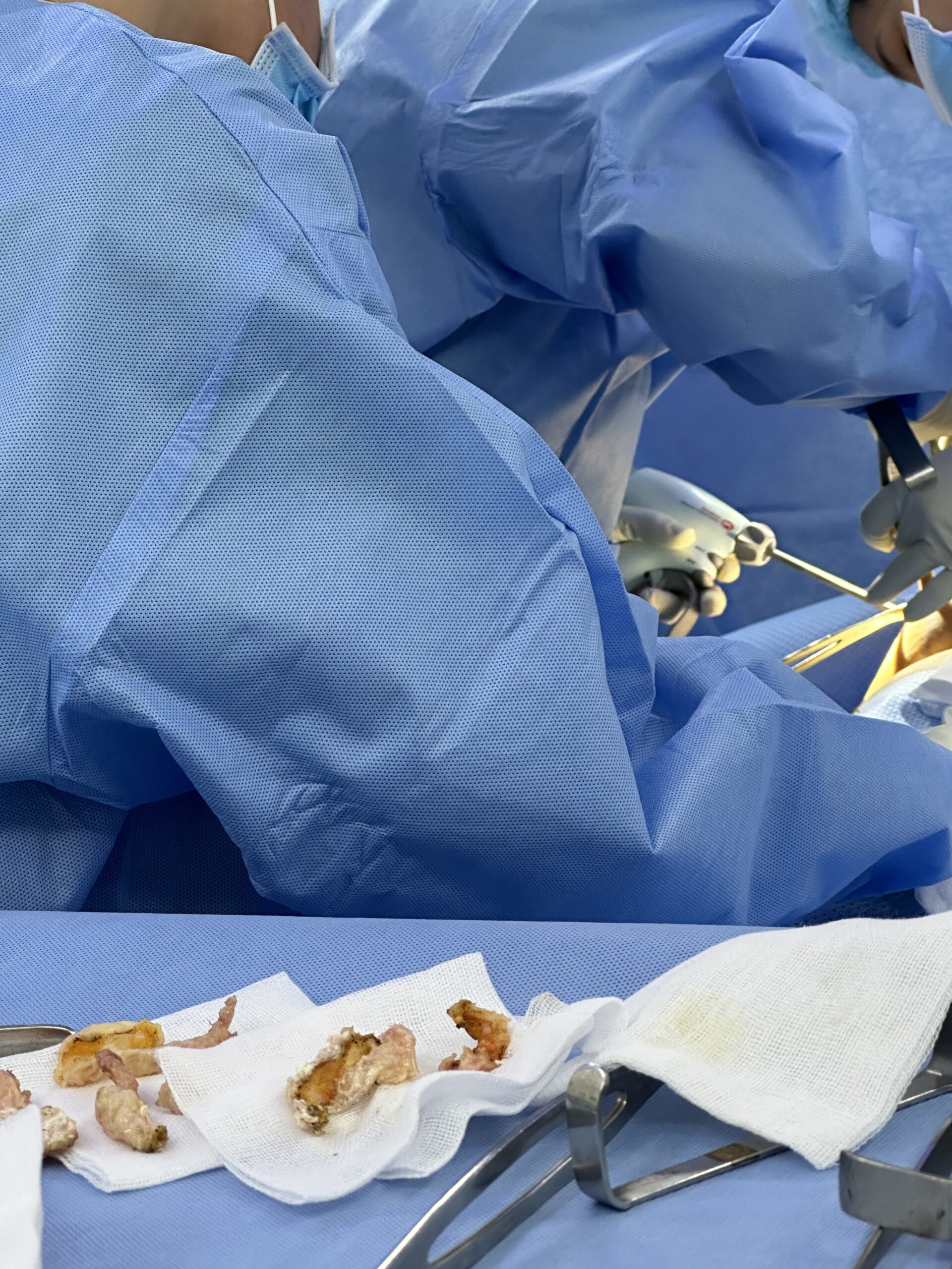
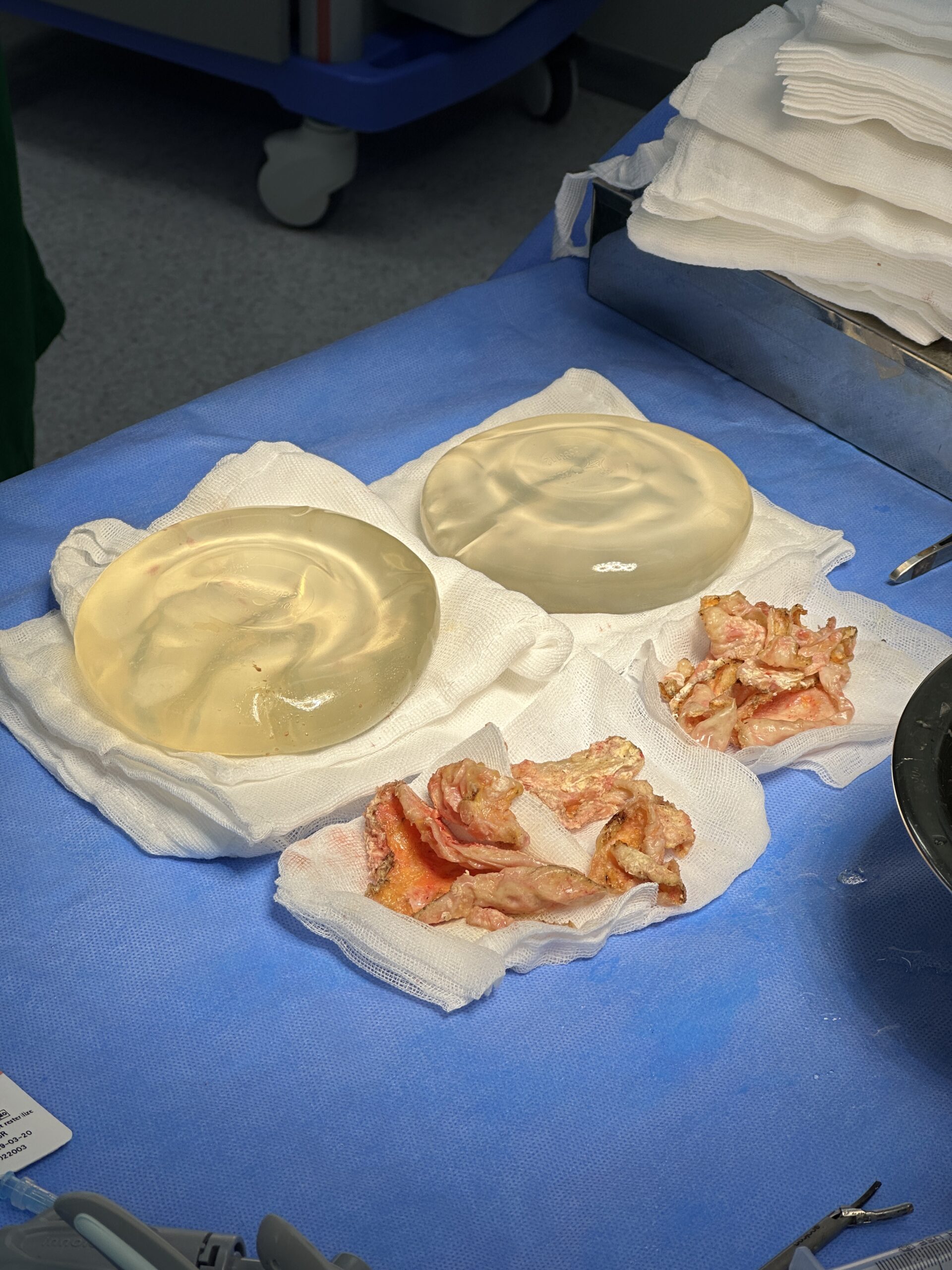
In some women, over a long period, calcium salts are laid down in the capsule. In a 1998 study of 404 capsules from silicone gel implants analysed between 1981 and 1996, Peters and colleagues described the calcification as white-grey plaques on the inner surface of the capsule, next to the implant. Under the microscope, every calcified capsule they examined showed two forms: globular mineral deposits on the surface and actual bone formation within the fibrous tissue.
With silicone gel implants, the implant itself is usually not the part that calcifies. Pagani and colleagues (2022) describe the deposits as small calcified plaques at the interface between the inner capsule and the implant surface. Saline implants behave differently: in a 2001 study of six saline implants removed after 7 to 23 years, Peters and colleagues found ivory-coloured deposits stuck to the front surface of the implant shell, with microscopic deposits in the adjacent capsule. Analysis of these deposits identified calcium apatite, a mineral of the same family as that found in bone.
How common it is and who is at risk
Most of what is known comes from older implant generations, because calcification takes many years to develop. The studies by Peters and colleagues give the most detailed figures available.
| Implant group (Peters et al., 1998) | Capsules with calcification | Time in place |
|---|---|---|
| First generation (1963–1972, Dacron patches) | 28 of 28 (100%) | mean 17.6 years (14–28) |
| Second generation (1973–1987) | 34 of 348 (9.8%) | mean 16.0 years (13–22) |
| Third generation (1987–1991) | 0 of 28 | mean 4.2 years (2–7) |
| Second generation, in place under 11 years | 0 of 294 | — |
| Second generation, 13 to 14 years | 33% | — |
| Second generation, 15 to 16 years | 45% | — |
| Second generation, 17 to 22 years | 57% | — |
Three risk factors stand out from these data:
- Time. None of the implants in place for less than 11 years showed significant calcification, and the proportion rose with longer duration.
- Implant generation. Older, thicker implants with Dacron patches were more likely to calcify, and the effect was not explained by duration alone.
- Rupture. Among second-generation implants in place for more than 12 years, calcification was found in 52.5% of ruptured implants and 10.0% of intact ones.
An earlier series by Peters and Smith (1995) found calcified capsules in 24 of 150 removed implants (16%). Every affected patient had calcification on both sides, and all 12 had Baker grade IV capsular contracture with pain as the main complaint. No implant in place for 10 years or less was calcified.
Modern implants differ from these devices. In a 2022 case report and literature review in Archives of Plastic Surgery, Pagani and colleagues noted that newer implant shells have made capsular calcification a rare finding. They also expected more cases in the coming years because of the number of women who now carry implants into older age. Their patient was 81 years old and had received smooth silicone implants more than 40 years earlier. Comparable large series for current cohesive gel implants are lacking, so the figures above describe older devices and should not be applied directly to recent ones.
Symptoms of a calcified breast implant capsule
Calcification often develops together with capsular contracture, which is why the symptoms overlap. The U.S. Food and Drug Administration (FDA) lists calcification and calcium deposits among the known complications of breast implants and describes them as hard lumps under the skin around the implant.
Changes that women commonly report include:
- a breast that feels firm or rock-hard, sometimes with an irregular edge;
- pain or tightness, particularly with a long-standing contracture;
- a change in breast shape, such as a high, round or distorted outline;
- a difference between the two sides, even though calcification usually affects both.
Symptoms and findings do not always match. Pagani and colleagues pointed out that some women with severe contracture report few symptoms, while others with milder changes are in considerable discomfort. Because the standard Baker grading does not include calcification, they proposed adding it to the grading system. Some calcified capsules are found incidentally, on a chest X-ray or a mammogram taken for another reason. The grading and symptoms of contracture itself are covered in our article on capsular contracture after breast augmentation.
How calcification shows on imaging
Calcium is dense on X-rays, so calcified capsules can show up on imaging done for another reason.
- Chest X-ray. In a pictorial review in Insights into Imaging (2011), Juanpere and colleagues showed capsule calcification as dense areas next to the implant on a plain chest film. In the 1995 series by Peters and Smith, one patient’s calcification was first seen on a routine chest radiograph.
- Mammography. The same review lists periprosthetic calcification among mammographic findings that are not specific to rupture. A calcified capsule does not by itself mean that the implant has failed.
- Ultrasound. Ultrasound shows the thickness of the capsule and can suggest rupture. In the case reported by Pagani and colleagues, it showed a thick fibrous capsule, an increased number of folds and signs of rupture inside the capsule.
- MRI. MRI remains the most accurate test for silicone implant rupture. Calcifications at the edge of the implant can appear as small points of altered signal, which the radiologist interprets alongside the rest of the scan.
The main concern on mammography is confusion with breast cancer. The FDA notes that calcium deposits around implants can be mistaken for cancer during mammography, resulting in additional surgery. Juanpere and colleagues illustrate the difference: calcification in the capsule lies next to the implant, whereas a cluster of fine microcalcifications inside the breast tissue is a separate finding that may need a biopsy, performed carefully to avoid damaging the implant. Pagani and colleagues add that calcification could interfere with tumour detection, which matters most for women whose implants were placed for reconstruction after breast cancer.
If you have implants, tell the mammography team how long they have been in place, and bring earlier images where possible so that the radiologist can compare them. Women with silicone implants are also advised by the FDA to have regular imaging even without symptoms, as described in our article on screening for silent rupture.
When a calcified capsule is removed
A calcified capsule is not an emergency in itself. The decision to remove it depends on symptoms, what imaging shows, the condition of the implant and what the patient wants for the breast afterwards.
A 2026 review in Aesthetic Plastic Surgery by Brown, graded by the journal as level V evidence, summarised what is known. It concluded that capsulectomy carries a higher complication rate than removing the implant alone, and that no published evidence documents harm from leaving a capsule in place after removal for benign reasons. It identified four indications supported by evidence:
- confirmed breast implant-associated anaplastic large cell lymphoma (BIA-ALCL);
- confirmed malignancy in the capsule;
- symptomatic capsular calcification with demonstrable impairment of mammography;
- grade III–IV capsular contracture where releasing the capsule (capsulotomy) is not enough.
Calcification appears in two of these four situations, directly or through the contracture that often accompanies it. Earlier authors reached similar conclusions from clinical experience: a 1989 case report by Young and colleagues recommended removing calcified capsules, and a 2006 letter by Barbosa and colleagues in Plastic and Reconstructive Surgery described capsulectomy as mandatory when capsular calcification is present.
| Situation | What is usually discussed |
|---|---|
| Hard, painful breast with a calcified capsule on imaging | Implant removal or exchange with total capsulectomy |
| Calcification that makes mammograms difficult to interpret | Capsulectomy, after discussion with the breast imaging team |
| Calcified capsule with a ruptured silicone implant | Removal of the implant and capsule, often as one piece where possible |
| Small, stable calcification found incidentally, no symptoms | Monitoring and imaging review, or removal if the patient prefers |
| Calcification alongside a new fluid collection or mass | Assessment first, including sampling, before planning surgery |
A thin capsule with no contracture, no calcification and an intact implant is a different situation, and leaving it in place can be reasonable. Our comparison of capsulectomy and capsulotomy for capsular contracture explains the options for capsules that are tight but not calcified.
What surgery for a calcified capsule involves
A heavily calcified capsule is stiff, can be thick and is sometimes firmly attached to the chest wall, so the operation is usually longer and involves more dissection than a straightforward implant exchange. A typical sequence:
- Step 1 — Assessment. History of the original surgery, implant type and age, symptoms and examination of both breasts.
- Step 2 — Imaging. Ultrasound, mammography where appropriate, and MRI when silicone rupture is suspected, to map the capsule and check the implant.
- Step 3 — Incision. The existing scar is used where possible.
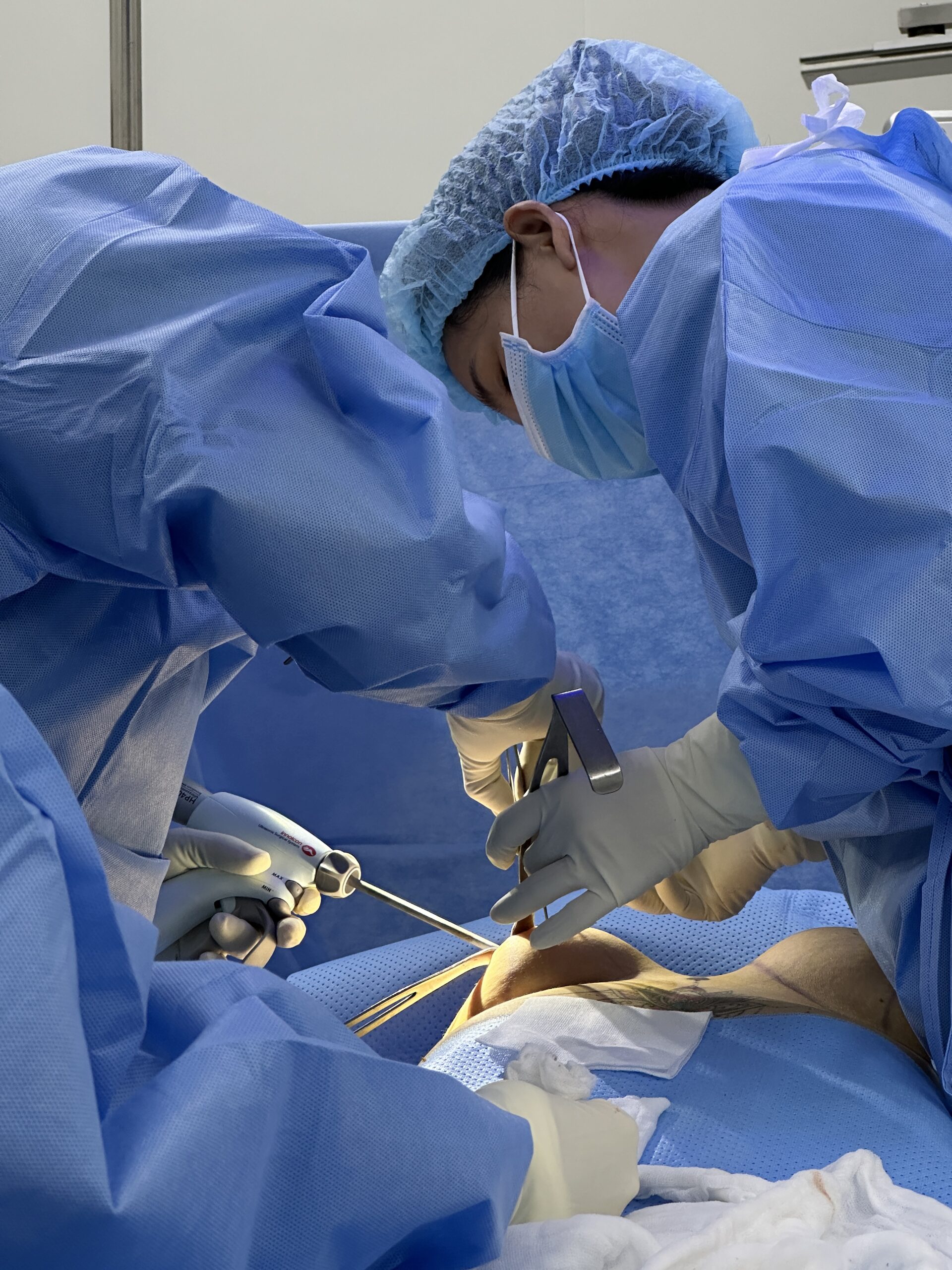
- Step 4 — Dissection and removal. The surgeon frees the capsule from the breast tissue and, where it lies on the ribs, from the chest wall. Where the implant is ruptured, the implant and capsule may be removed together to avoid spreading silicone in the pocket. In the case reported by Pagani and colleagues, the calcification was so extensive that the specimen could not be removed without breaking it.
- Step 5 — Pathology. The capsule can be sent for histological examination, which confirms calcification and checks for other findings.
Dissection over the chest wall carries a small risk of injury to the underlying tissues, and recovery is usually longer than after a simple implant exchange because more tissue has been disturbed. After many years of contracture, the breast may look smaller or flatter once the implant and capsule are out. Some women choose replacement or a breast lift, while others prefer no implant. Our article on breast implant removal without replacement describes what to expect if no new implant is placed.
Assessment and capsulectomy for calcified capsules in Vietnam
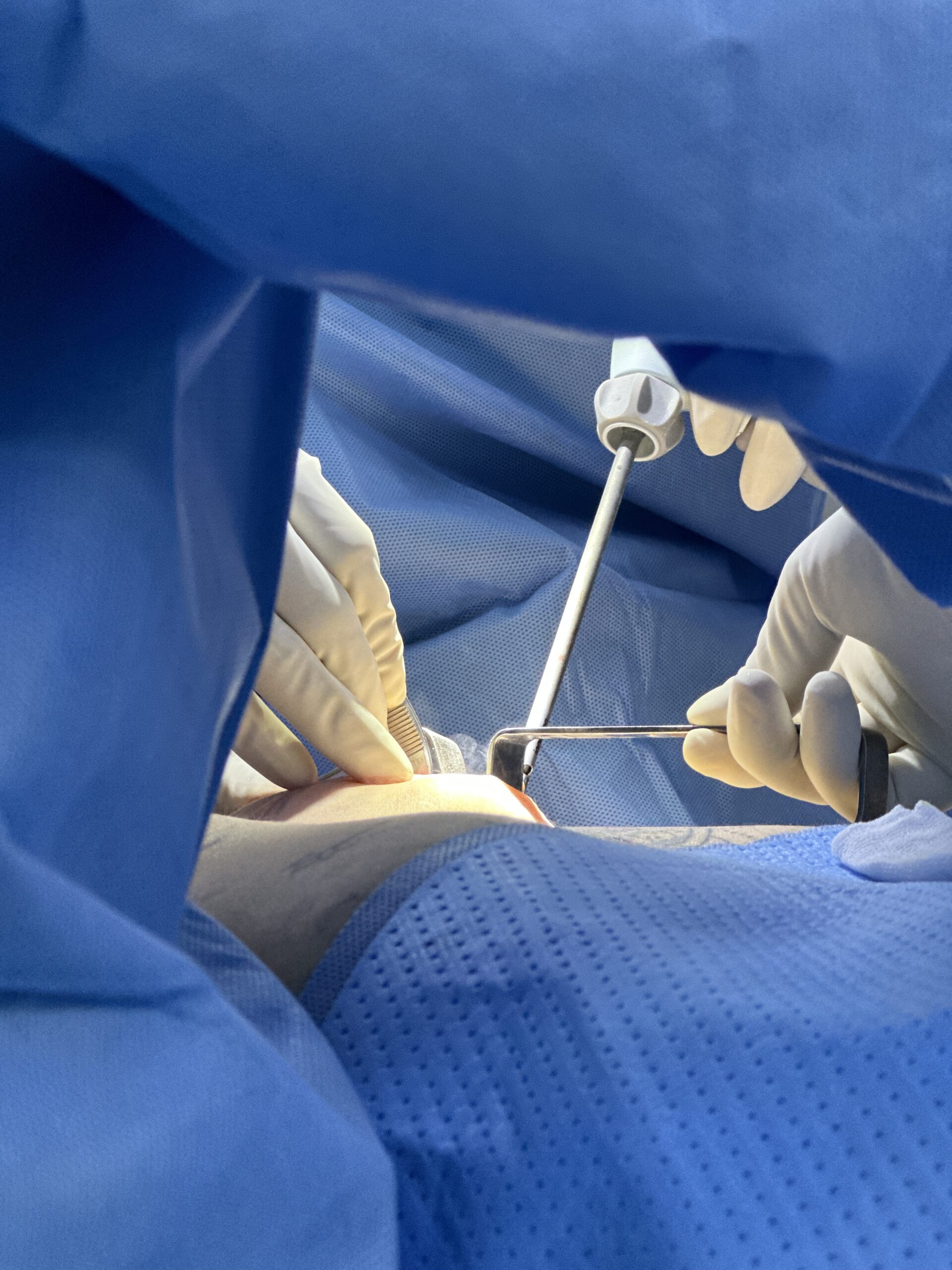
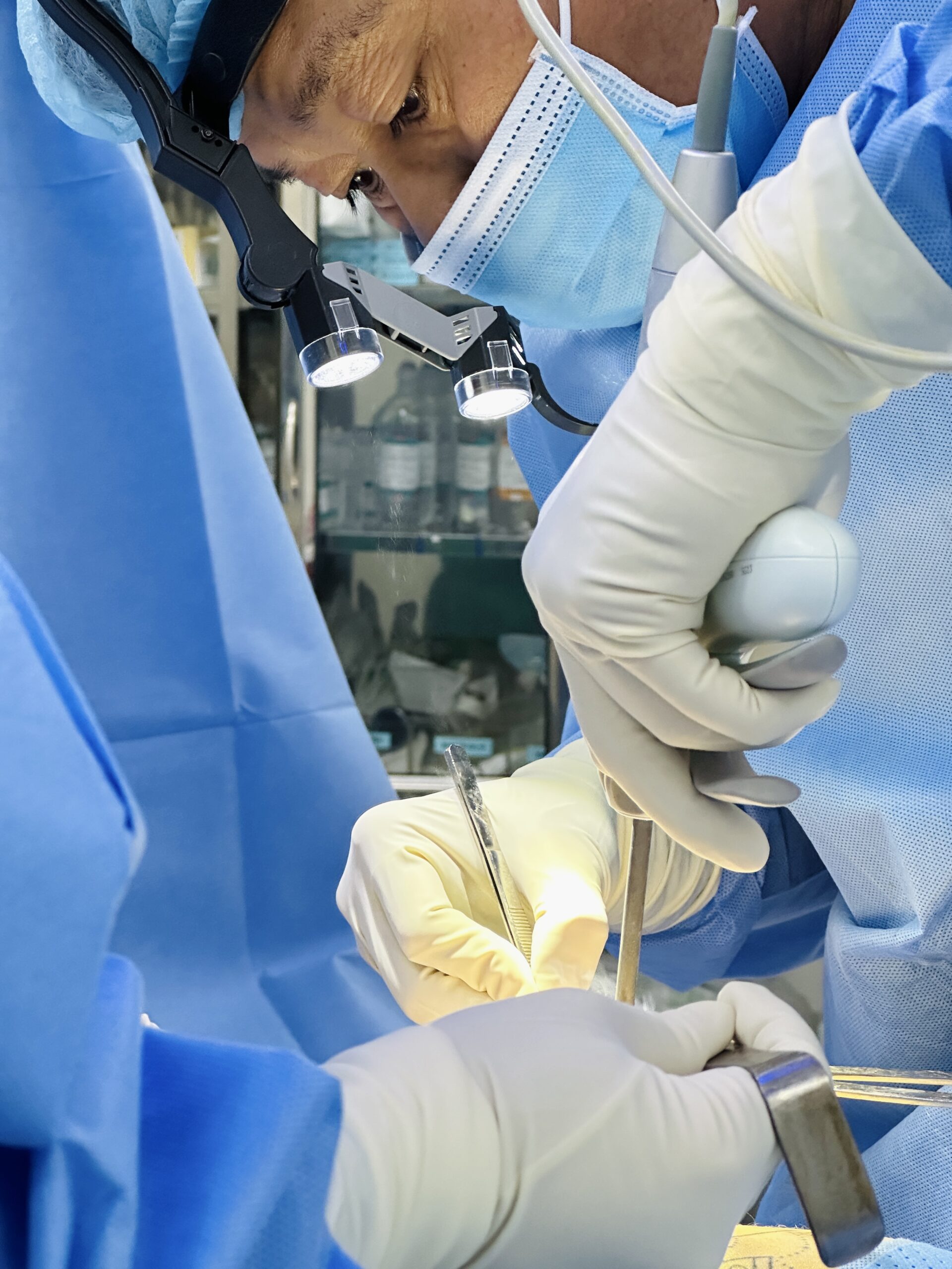
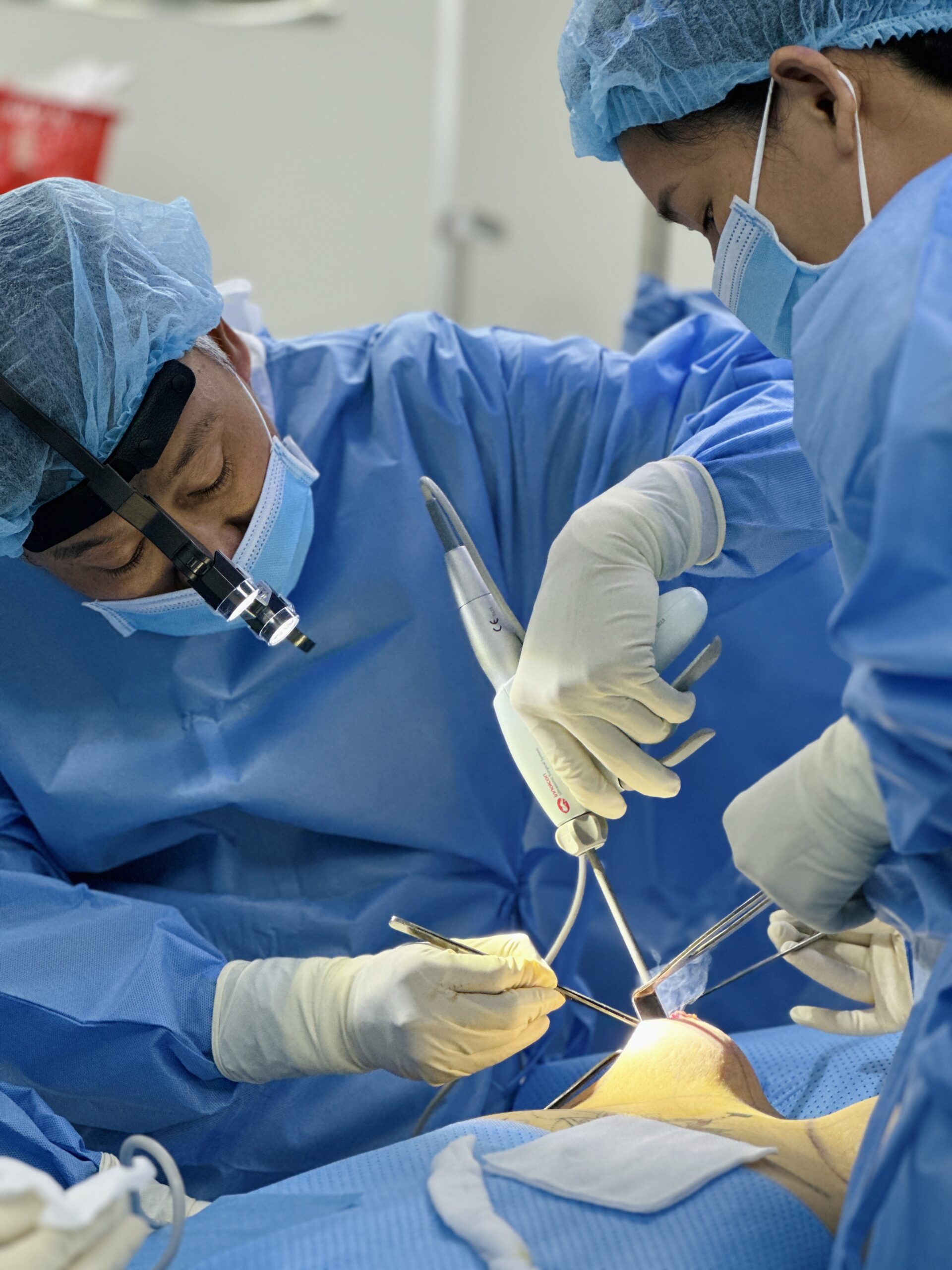
At Dr. Breast X, MSc, MD Ho Cao Vu performs breast implant removal with partial, total or intact capsulectomy at the Department of Surgery, Nam Sai Gon International General Hospital in Ho Chi Minh City. The extent of capsule removal is planned from the history, examination and imaging, with ultrasound first and MRI where silicone rupture is suspected. A thickened, calcified or contracted capsule is one of the situations in which a total capsulectomy is considered.
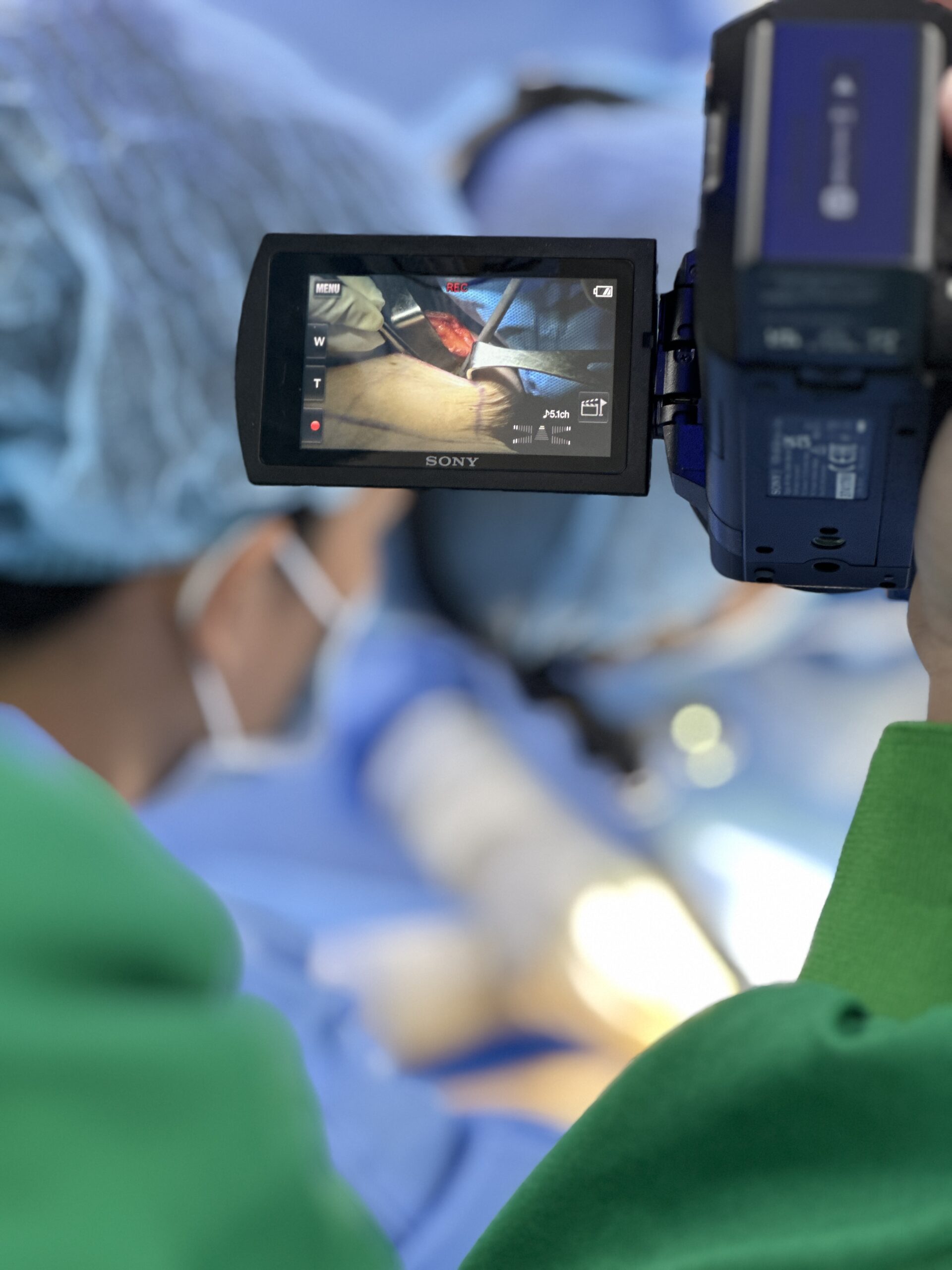
Dissection is performed with an ultrasonic surgical scalpel, which divides tissue and coagulates small vessels in the same pass. Our video library includes a recorded capsulectomy of a calcified capsule 20 years after saline implant placement. For women travelling from abroad, costs and the steps of planning are set out in our guide to breast implant removal in Vietnam.
Frequently asked questions
What does it mean if my breast implants are calcified? It usually means that calcium deposits have formed in the scar capsule around the implant, rather than in the implant itself. It is most often found around implants that have been in place for many years, and it is frequently linked with capsular contracture.
Is a calcified capsule cancer? No. Calcification of the capsule is a reaction of the scar tissue, not a tumour. The concern is that it can make mammograms harder to read or be mistaken for a suspicious finding, so new changes in the breast tissue still need proper assessment.
Can saline implants calcify? Yes. Calcification has been described around saline implants, where deposits form mainly on the front surface of the implant shell and in the adjacent capsule.
Does a calcified capsule mean the implant has ruptured? Not necessarily. Calcification was more common around ruptured older implants in the Peters series, but it also occurs around intact ones. Imaging, usually MRI for silicone implants, is needed to check the implant.
Conclusion
A calcified breast implant capsule is a long-term change in the scar tissue around an implant, found mainly with older devices that have been in place for well over a decade. It can cause a hard, painful breast, it shows on mammograms and X-rays, and it can be confused with a suspicious finding. Current evidence supports capsulectomy when calcification causes symptoms or impairs breast imaging, while a small, stable deposit can be monitored. The plan is made from examination and imaging.
References
- U.S. Food and Drug Administration — Risks and Complications of Breast Implants (content current as of 15 December 2023) — https://www.fda.gov/medical-devices/breast-implants/risks-and-complications-breast-implants
- Peters W, Pritzker K, Smith D, et al. Capsular calcification associated with silicone breast implants: incidence, determinants, and characterization. Annals of Plastic Surgery 1998;41(4):348–360 — https://doi.org/10.1097/00000637-199810000-00002
- Peters W, Smith D. Calcification of breast implant capsules: incidence, diagnosis, and contributing factors. Annals of Plastic Surgery 1995;34(1):8–11 — https://doi.org/10.1097/00000637-199501000-00002
- Peters W, Smith D, Lugowski S, Pritzker K, Holmyard D. Calcification properties of saline-filled breast implants. Plastic and Reconstructive Surgery 2001;107(2):356–363 — https://doi.org/10.1097/00006534-200102000-00010
- Pagani A, Aitzetmüller MM, Larcher L. A Forgotten Entity following Breast Implant Contracture: Does Baker Need a Change? Archives of Plastic Surgery 2022;49(3):360–364 — https://doi.org/10.1055/s-0042-1744409
- Juanpere S, Perez E, Huc O, et al. Imaging of breast implants — a pictorial review. Insights into Imaging 2011;2(6):653–670 — https://doi.org/10.1007/s13244-011-0122-3
- Brown T. Capsulectomy Following Breast Implant Removal: Evidence-Based Indications and a Framework for Clinical Decision-Making. Aesthetic Plastic Surgery 2026 (published online 15 October 2026) — https://doi.org/10.1007/s00266-026-06365-2
- Young VL, Bartell T, Destouet JM, Monsees B, Logan SE. Calcification of breast implant capsule. Southern Medical Journal 1989;82(9):1171–1173 — https://doi.org/10.1097/00007611-198909000-00029
- Barbosa MV, Nahas FX, Ferreira LM. Capsulectomy: a mandatory procedure in the presence of capsular calcification. Plastic and Reconstructive Surgery 2006;117(5):1654–1655 — https://doi.org/10.1097/01.prs.0000208921.66204.85


