Silicone in Lymph Nodes After Breast Implants: Symptoms, Whether It Is Dangerous and What Removal Can Change
MSc, MD Ho Cao Vu
Medical practice licence No. 028475/BYT-CCHN, issued by the Ministry of Health of Vietnam on 28 June 2018. Specialist in General Surgery and Plastic & Aesthetic Surgery, with over 15 years of experience in plastic and aesthetic surgery and over 20 years of general surgical experience in benign breast disease, breast cancer, abdominal wall and abdominal surgery at Cho Ray Hospital.
Updated November 2026 — cross-checked against a 2025 ten-year imaging study of 1,217 women with silicone implants and a 2026 study of 299 surgically confirmed implant ruptures.
An ultrasound report mentions a “snowstorm” in the armpit, or a small lump appears there years after breast augmentation. One possible explanation is silicone lymphadenopathy: tiny silicone particles from the implant have collected in nearby lymph nodes. It often causes no symptoms and is usually benign, but it can point to a ruptured implant and can look like cancer on a scan. Below, we look at how common it is, how it is confirmed, when a biopsy is needed and what implant removal can and cannot change.
The short answer: is silicone in the lymph nodes dangerous?
Silicone in a lymph node is a foreign body reaction, not a cancer. In a series by Desai and colleagues (Miami, 2025), 36 of 52 women had no symptoms, none of the 8 women who had a biopsy had cancer, and no one needed the nodes removed. A 2022 case report and literature review by Elahi and colleagues (Lausanne) concludes that these nodes need no follow-up or special treatment unless they cause symptoms or interfere with checking for cancer recurrence.
That reassurance has two limits. The finding may mean the implant has ruptured, and an enlarged node should not be assumed to be silicone, particularly after breast cancer.
How silicone reaches the lymph nodes
The FDA describes three patterns of rupture in a silicone gel implant. Generally the gel stays inside the scar tissue around the implant (intracapsular rupture). If it moves beyond that scar tissue, the rupture is extracapsular. If it travels to more distant areas, the FDA calls it gel migration. Our article on silent breast implant rupture explains why many of these ruptures go unnoticed.
Rupture is not the only route. The Desai paper explains that silicone is often carried by macrophages, immune cells that clear foreign material, and that the resulting foreign body reaction enlarges the node. Berben and colleagues (Maastricht, 2025) note that silicone particles can also pass through an intact shell.
You may see these terms in a report:
- Silicone lymphadenopathy: lymph nodes enlarged by silicone. Also called silicone adenopathy or adenitis.
- Siliconoma or silicone granuloma: a firm lump formed by the inflammatory reaction to silicone. Elahi and colleagues note that these can occur in the breast, the skin and the nodes.
- Gel bleed: microscopic passage of silicone through a shell that has not torn.
- Snowstorm appearance: the speckled, bright pattern silicone produces on ultrasound.
How common is it?
Three recent studies asked different questions, so they cannot be compared directly.
| Study | Who was studied | Silicone in lymph nodes | What it adds |
|---|---|---|---|
| Berben and colleagues, 2025 (Netherlands) | 1,217 women with silicone implants; imaging reports from 2010 to 2020 | 71 women (5.8%): 24 of 160 with rupture (15%), 47 with implants that looked intact (4.4%) | The only one of the three to measure how common it is across women with implants |
| Bresnick, 2026 (United States) | 260 women, 299 ruptured smooth fourth-generation implants confirmed at surgery | 46 of 299 implant sides (about 15%) | Odds rose with the age of the implant |
| Desai and colleagues, 2025 (United States) | 52 women already diagnosed with silicone lymphadenopathy | Rupture of a current or earlier implant in 46; no rupture in 6 | Found a median of 12.5 years after implant placement |
In the Bresnick study, each extra year of implant age was linked with about 13% higher odds of silicone in the armpit nodes, while extracapsular rupture added only a modest independent effect. Nodes were affected in 34 of 244 intracapsular ruptures and 12 of 55 extracapsular ruptures, so a rupture contained within the capsule does not rule them out. Implant size and manufacturer made no significant difference.
In the Berben study, 17 of the 47 women with apparently intact implants had no earlier implant and no known rupture of one, which suggests that silicone can reach the nodes without any rupture. The authors add that many cases are found by chance, so the true figure may be higher. Both studies come from one centre, and Bresnick’s covers only ruptures that went to surgery.
Symptoms: what women actually notice
Most women notice nothing. The Desai series reported:
- No symptoms: 36 of 52 women (69.3%).
- Painful enlarged nodes: 10 women (19.2%).
- Painless enlarged nodes: 4 women (7.7%).
- Both painful and painless nodes: 1 woman (1.9%).
- General symptoms: fever, chills, weight loss, weakness or fatigue in 1 woman.
The armpit was the main site, in 45 of the 52 women (86.5%). Two had nodes only along the breastbone (internal mammary nodes), and 5 had more than one region involved. Fifteen women (28.9%) had nodes on both sides. Elahi and colleagues describe a rarer pattern in which silicone appears in the armpit on the opposite side.
The FDA adds that a rupture occasionally shows as a smaller breast, a change in implant shape, hard lumps, unevenness, pain, tingling, swelling, numbness or burning. Whether general symptoms such as fatigue come from silicone is unsettled: the Desai paper notes they can be hard to separate from breast implant illness.
How it is confirmed: ultrasound, MRI and biopsy
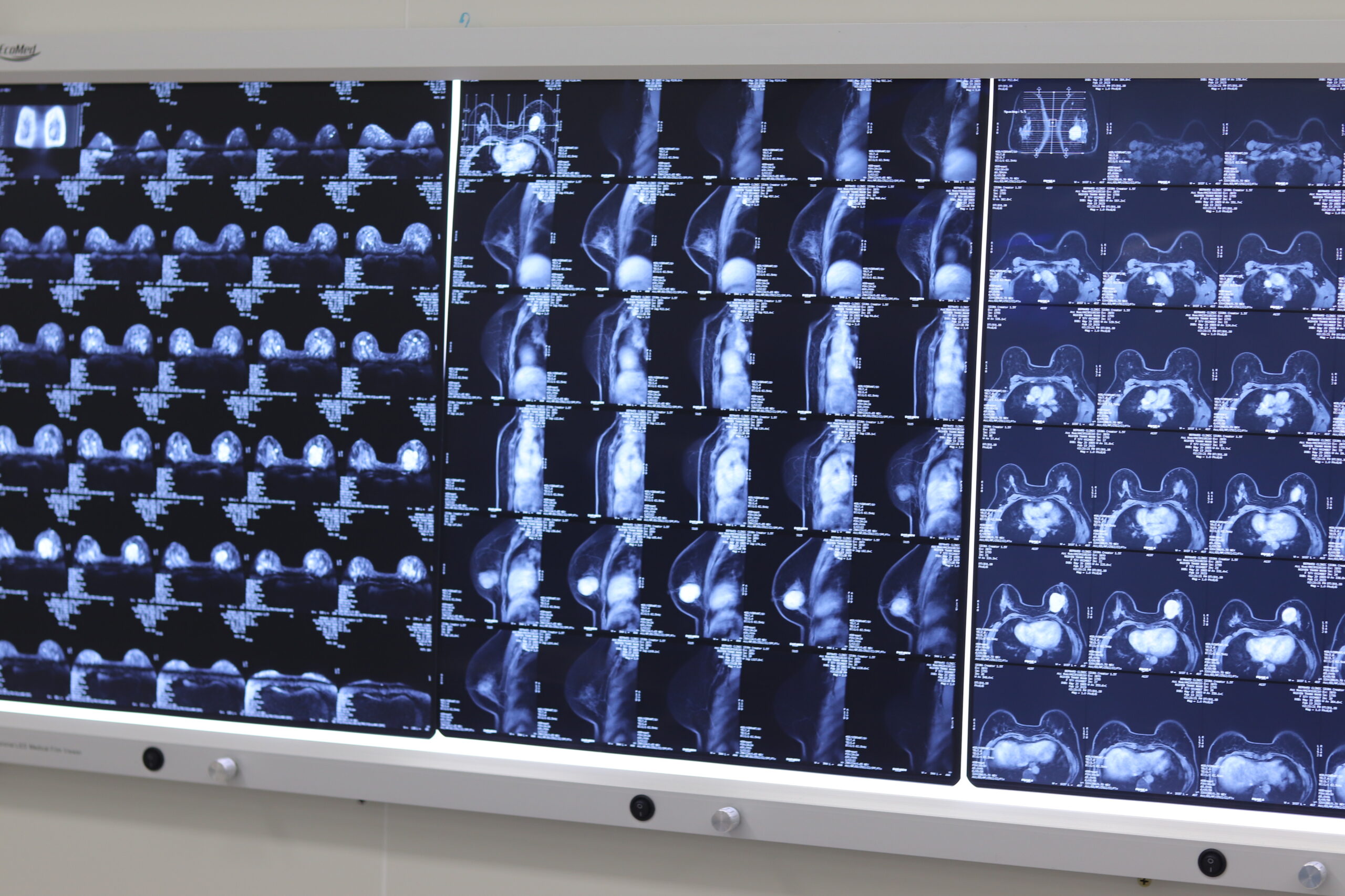
Ultrasound is the usual test for the nodes themselves. In the Desai series it detected the finding in 42 of 52 women (80.7%), mammography in 35 (67.3%), MRI in 9 (17.3%) and CT in 2, with some women having more than one test. Both Elahi and Berben state that ultrasound is more sensitive than MRI for silicone in lymph nodes. The Desai paper cites a reported sensitivity of 87.5% and specificity of 100% for the snowstorm sign, and suggests that a low-risk woman with painless nodes and this pattern may not need a biopsy.
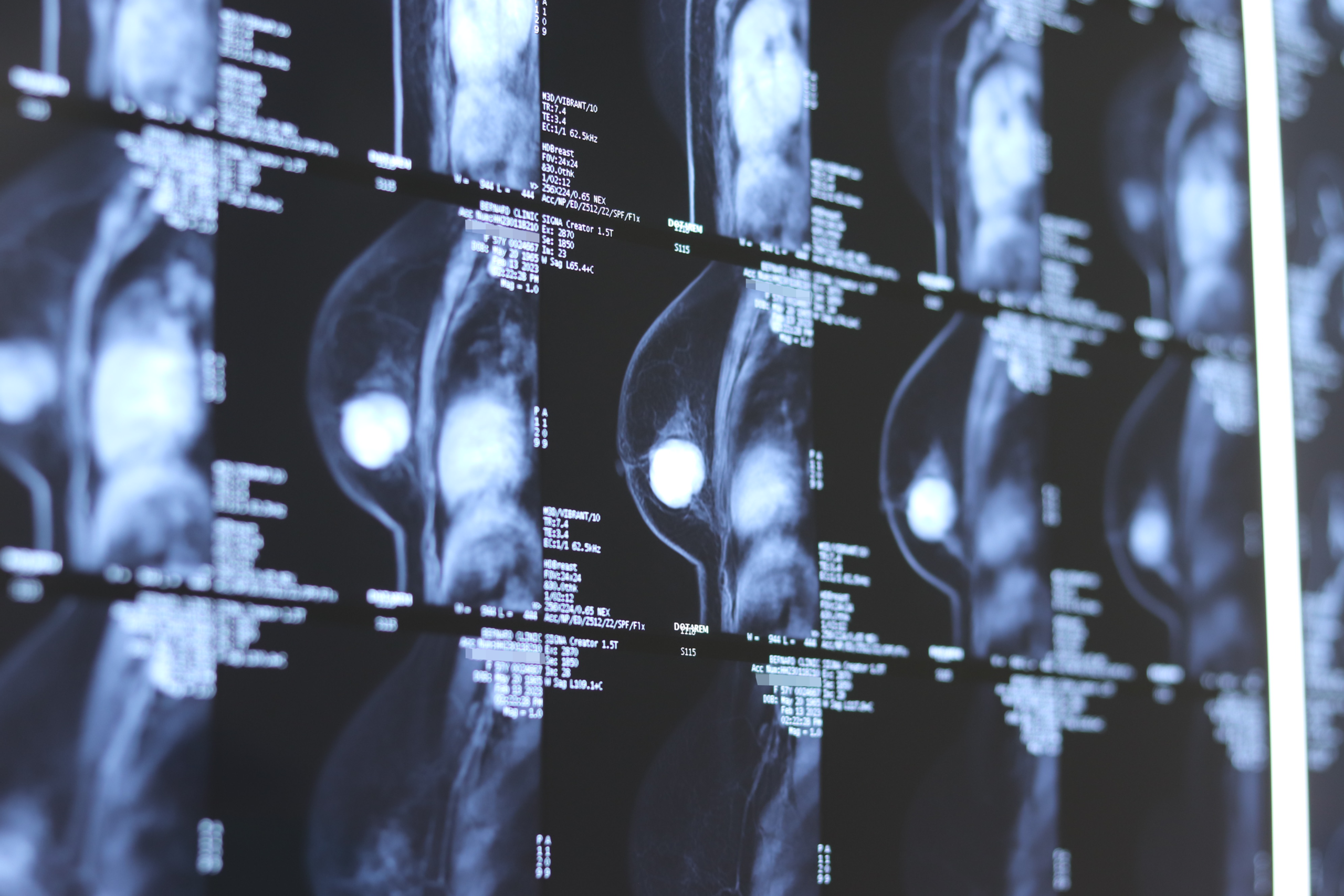
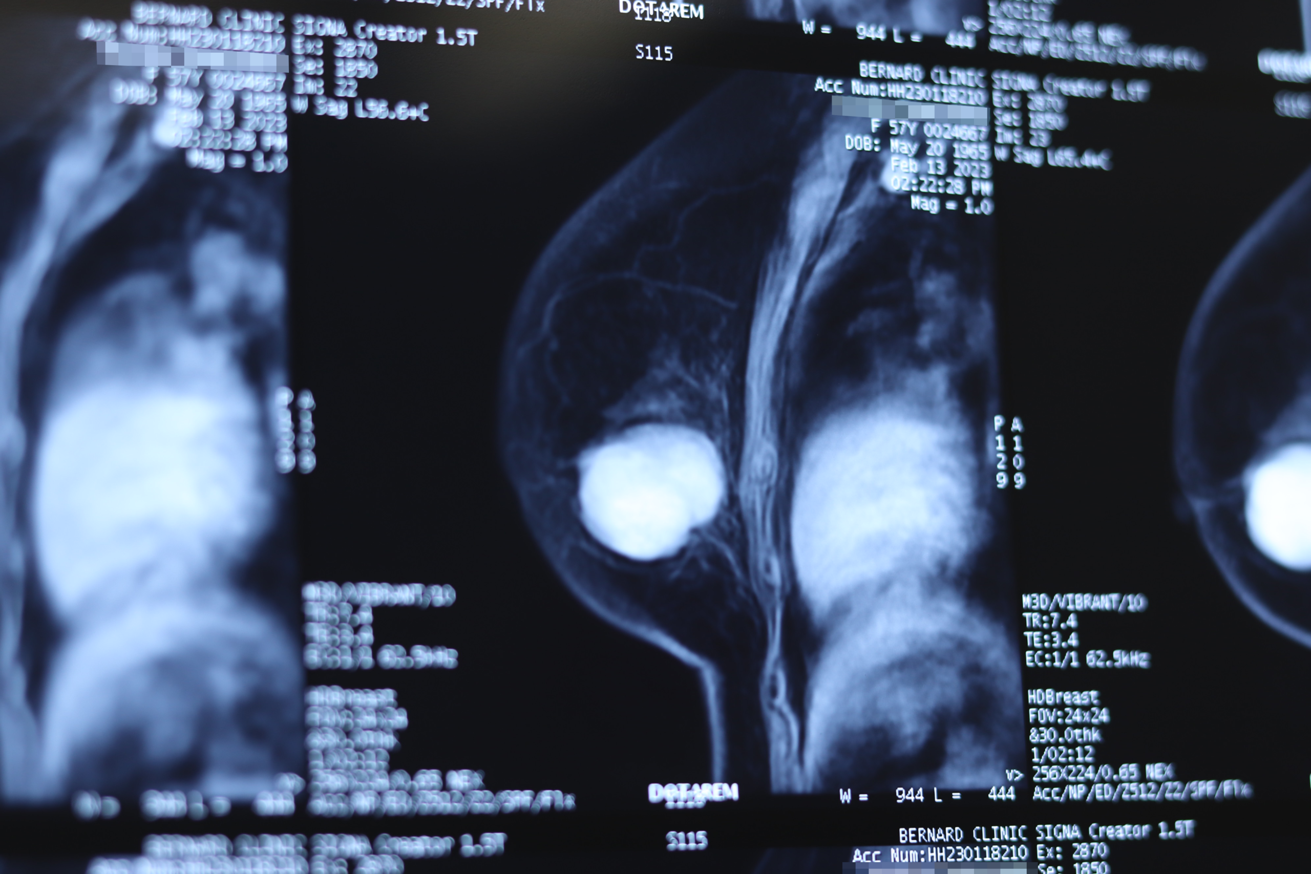
MRI answers a different question. The FDA calls it the most effective method for detecting a silent rupture and describes ultrasound as an acceptable alternative for screening women without symptoms. In the Berben study, among implants later checked at surgery, MRI detected 91.7% of ruptures and ultrasound 76.2%. The authors caution that these subgroups were small. So a woman with silicone in her nodes may need both tests: one for the nodes and one for the implant.
PET-CT can mislead. Berben and colleagues warn that silicone nodes can appear active on this scan and be taken for cancer.
Biopsy is kept for uncertain cases. In the Desai series, 8 of 52 women (15.4%) had one because of suspicious imaging. Seven showed benign silicone changes, one showed other findings and none showed cancer. Elahi and colleagues note that for armpit nodes, a fine needle aspiration under ultrasound guidance is sufficient.
Could it be cancer? When not to assume it is silicone
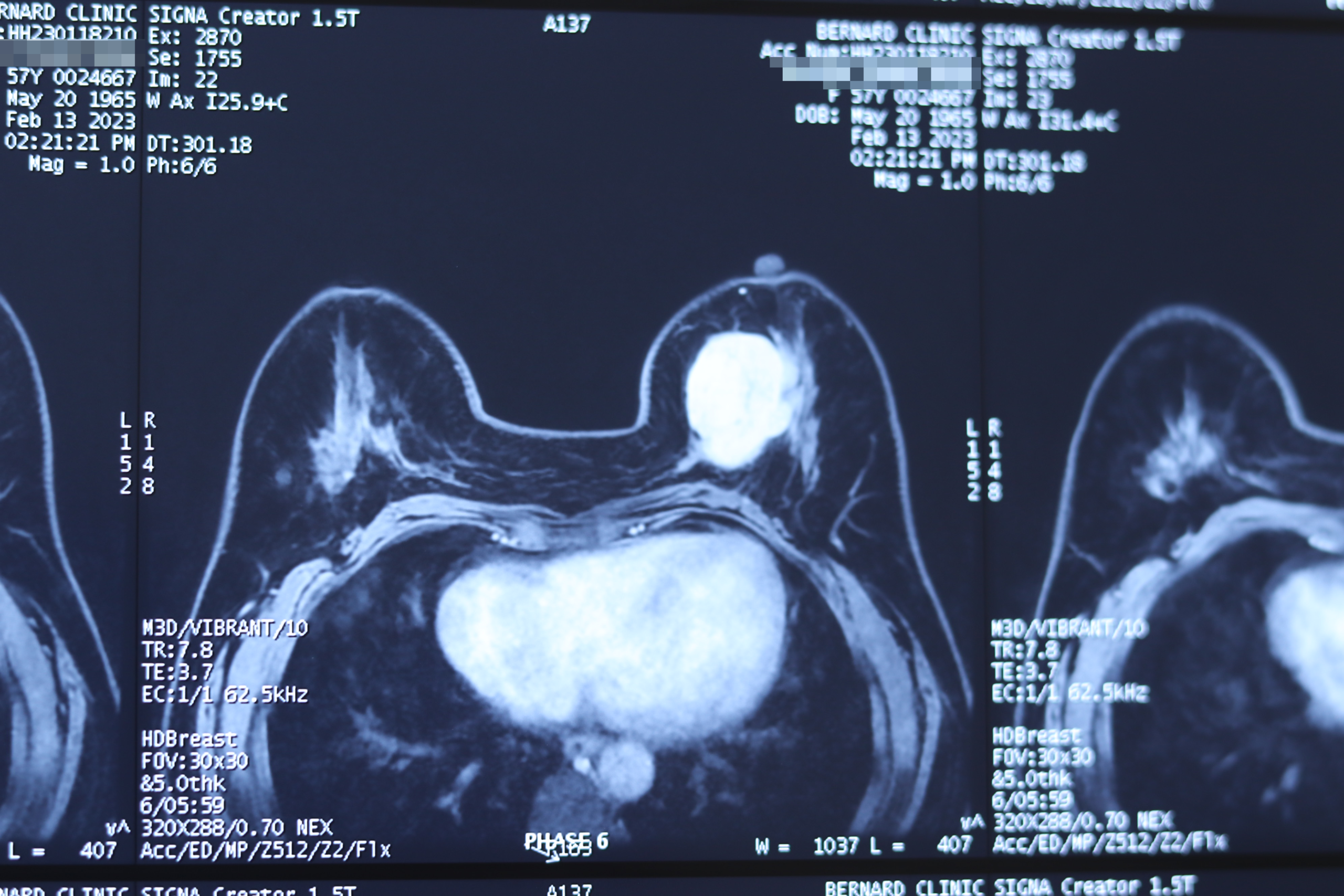
The FDA states it has not detected an association between silicone gel implants and breast cancer. The studies cited here were not designed to measure long-term cancer risk from silicone in the nodes. The practical concern is different: silicone can sit alongside a cancer, or hide one.
- A history of breast cancer changes the assessment. In the Desai series, 8 of 52 women had a history of breast cancer, and in 2 a recurrence was diagnosed when the silicone nodes were found. The authors call evaluation essential in this group.
- Silicone can obscure imaging. Chua and colleagues (Singapore, 2025) describe one woman with injected liquid silicone, not implants, whose breast lump was attributed to silicone for several years. When it grew and became painful, excision showed an 80 mm invasive carcinoma. One case gives no rate, but the lesson applies to any lump that changes.
- Persistent swelling around an implant has its own workup. The FDA advises talking to your surgeon about persistent swelling, pain or other changes around the implant because of BIA-ALCL, a rare lymphoma. Our article on BIA-ALCL covers the symptoms.
Ask for prompt assessment if a lump in the breast or armpit is new, growing or persistently painful, or if you have had breast cancer.
Do silicone lymph nodes have to be removed?
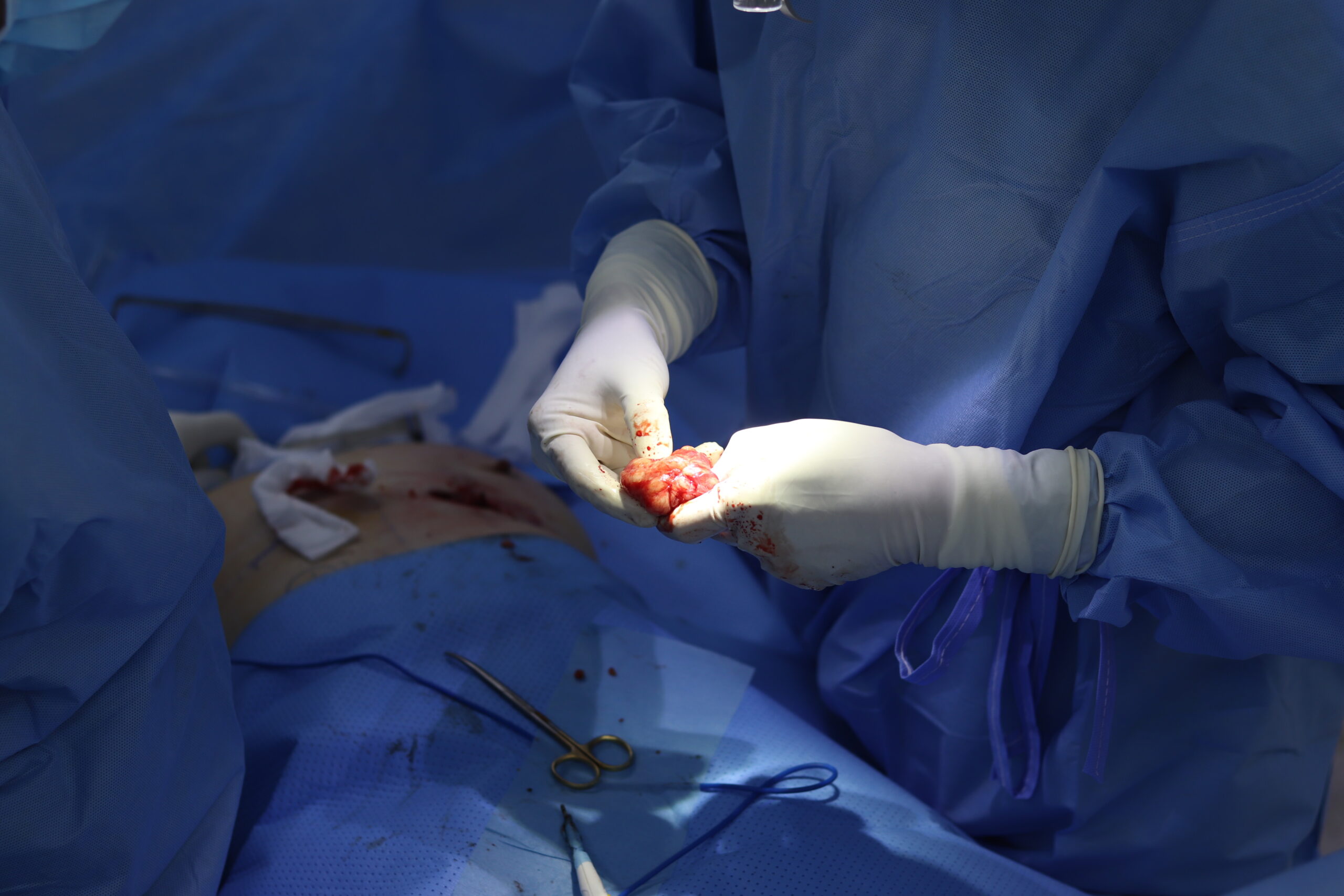
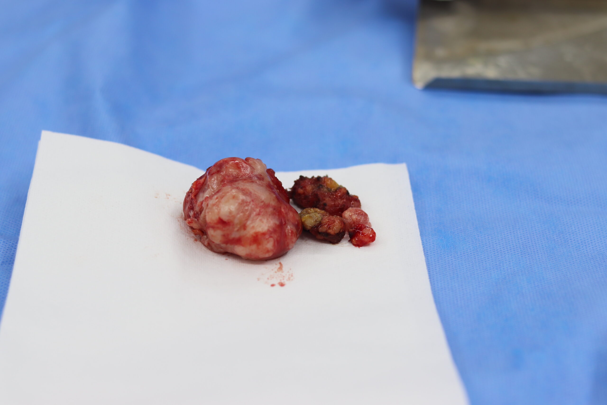
Usually not. In the Desai series, no woman was treated by removing the nodes alone. The authors advise against removing nodes that cause no symptoms because of the risk of lymphedema, a lasting swelling of the arm. They say excision may be considered for painful nodes, but earlier reports are mixed: some women improved, while others had persistent symptoms or recurrence.
Elahi and colleagues report no specific follow-up at their Lausanne centre once a biopsy has shown silicone. Their own patient, who had no symptoms, was observed, and her nodules were unchanged on MRI at 6 months.
Does implant removal clear silicone from the nodes?
The evidence says not reliably. Desai and colleagues state that removing implants does not cure silicone lymphadenopathy or prevent it from developing later. Their series points the same way: in 28 of the 46 women with a rupture, it was an earlier implant, since removed or exchanged, that had ruptured. The FDA also warns that silicone gel may be difficult to remove after a rupture, and the Desai paper notes that removal itself carries some risk of spreading silicone.
So the decision is usually made on the condition of the implant:
- Rupture confirmed. Across the Desai series, 13 women had explantation and 21 an implant exchange, counting surgery before or at diagnosis. Another 18 had no surgery, 6 of them with intact implants. Bresnick saw some untreated ruptures progress on repeat imaging, to extracapsular spread or to silicone in the nodes. He encourages timely management, while accepting that his study cannot prove cause.
- Implant intact. The finding may reflect gel bleed. In the Berben study, 16 of the 47 women in this group had their implants removed. No study we read shows that removal helps this group, so the decision rests on symptoms and your own wishes.
If you have surgery, the plan also covers the capsule. Our article on en bloc, total and partial capsulectomy explains what changes when silicone has already left it.
Follow-up after the diagnosis
For women without a breast cancer history, Desai and colleagues see no need for monitoring beyond standard breast screening. Women with implants still in place continue rupture screening: the FDA’s schedule, as cited in the Desai paper, starts 5 to 6 years after implantation and repeats every 2 to 3 years.
Silicone nodes may remain visible after removal, so tell every radiologist about them, keep earlier images and reports, and mention when any implant was removed. Our article on mammograms with breast implants explains what to tell the imaging clinic.
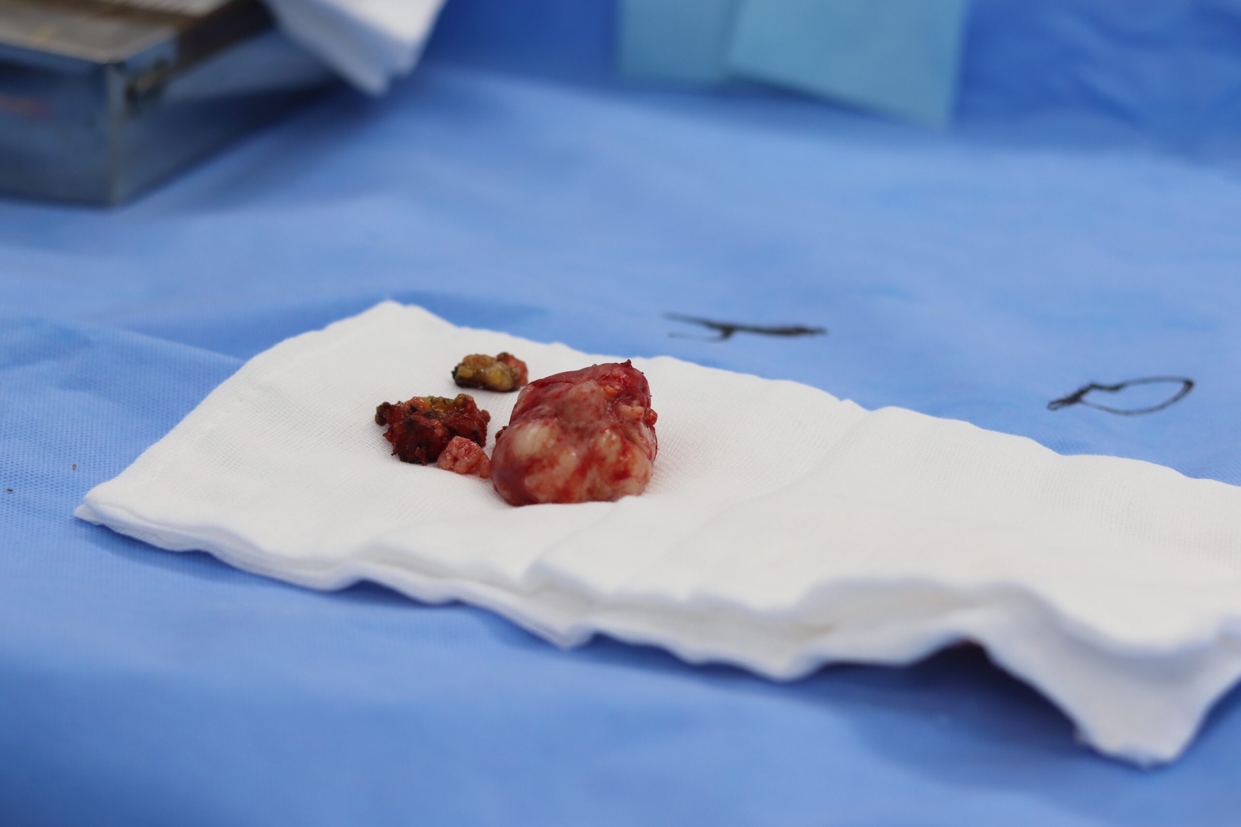
How the decision is made at consultation
- Step 1 — Take the history. Implant age and type, any earlier rupture or exchange, and any breast cancer.
- Step 2 — Review the imaging. Ultrasound of the breast and armpit, and MRI if the state of the implant is unclear.
- Step 3 — Decide whether a biopsy is needed. It is considered for suspicious imaging, persistent symptoms or a cancer history.
- Step 4 — Decide about the implant. Keep it under surveillance, exchange it or remove it, based on its condition and your goals.
- Step 5 — Agree the follow-up. Breast screening, rupture screening for any implant that stays, and what to report early.
Silicone in lymph nodes and implant removal in Vietnam
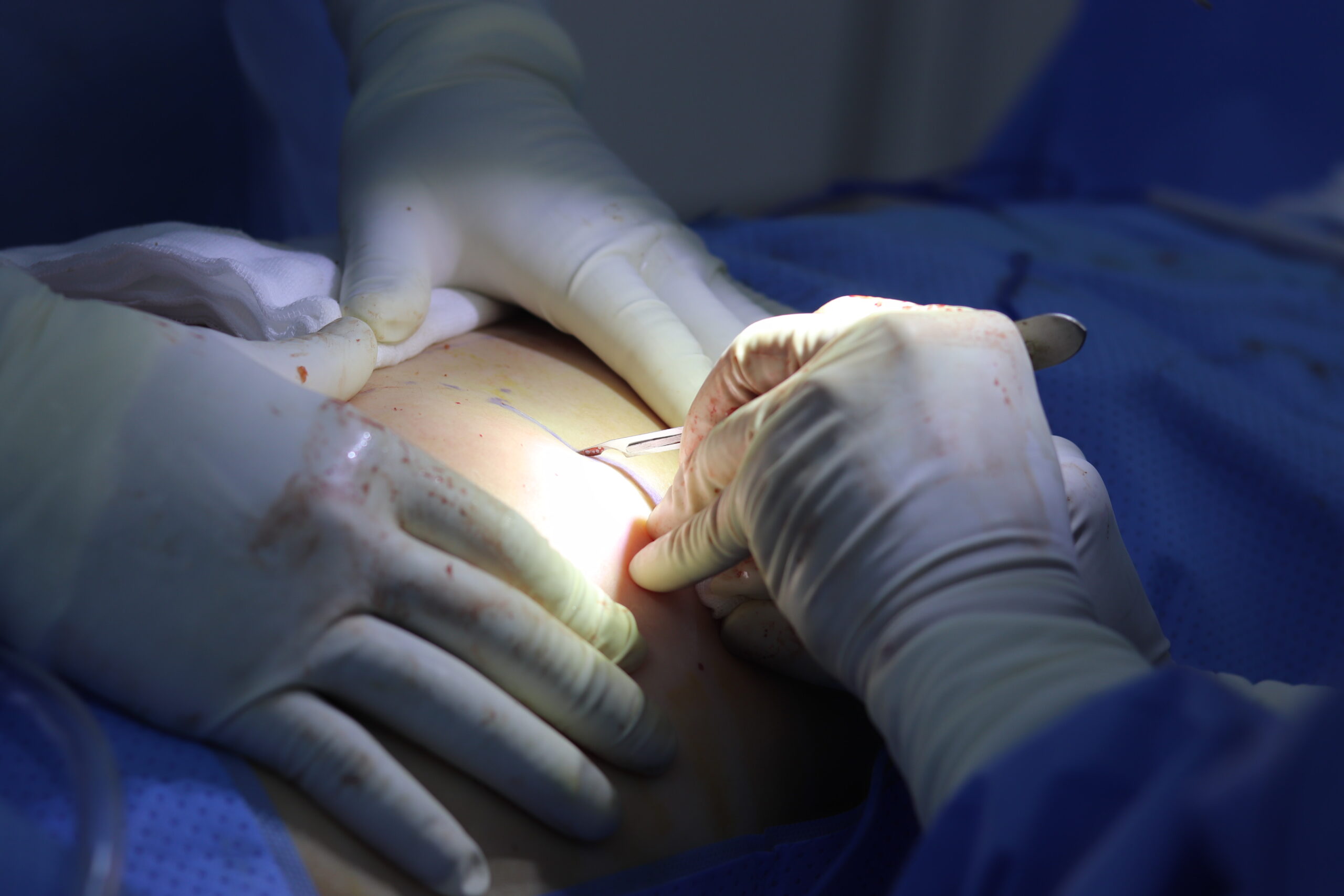
Consultations and preoperative assessments are provided at Dr. Breast X. Surgery is performed directly by MSc, MD Ho Cao Vu at the Department of Surgery, Nam Sai Gon International General Hospital in Ho Chi Minh City, under laryngeal mask airway (LMA) anaesthesia and with an ultrasonic surgical scalpel. How any lymph node finding is handled is discussed individually at consultation.
If you live overseas, send your imaging reports and the images themselves before you travel, including any armpit ultrasound and any earlier biopsy result. Our guide to planning from overseas lists what is useful. Planning figures for routine removal are in our guide to breast implant removal in Vietnam. A lymph node workup may change the plan, so the length of stay is confirmed at consultation.
Frequently asked questions
Is silicone in the lymph nodes dangerous? It is usually benign. In the Desai series, most women had no symptoms and no biopsy showed cancer. The implant may still need checking for rupture.
Can silicone be removed from lymph nodes? Nodes can be removed surgically, but this is not routine. Desai and colleagues advise against it when there are no symptoms because of the risk of lymphedema, and results for painful nodes are mixed.
Will the lymph nodes go back to normal after explant? This cannot be promised. Desai and colleagues state that removing implants does not cure the condition, so tell your radiologist about the nodes.
Can it happen if my implant is not ruptured? Yes. In the Berben study, 47 women with implants that looked intact on imaging had silicone in their nodes, though some had earlier implants that had ruptured. Silicone can also pass through an intact shell.
Conclusion
Silicone in the lymph nodes is usually a benign finding, seen in about 6% of women with silicone implants in one ten-year study and about 15% of ruptured implants in two. Most women have no symptoms, and the nodes rarely need removing. The real questions are whether the implant has ruptured and whether a lump could be something else, especially after breast cancer. Implant removal does not reliably clear the nodes, so any decision about surgery rests on the implant, your symptoms and your goals.
References
- Berben JA, Heuts EM, van Nijnatten TJA, van der Hulst RRWJ. Prevalence of Silicone Lymphadenopathy in Women with Breast Implants: A single-center retrospective study. JPRAS Open 2025;44 — https://doi.org/10.1016/j.jpra.2025.01.016
- Bresnick SD. Silicone Axillary Lymphadenopathy Associated With Rupture of Fourth-Generation Cohesive Silicone Breast Implants Following Breast Augmentation. Aesthetic Surgery Journal 2026;46(9):1007–1011 — https://doi.org/10.1093/asj/sjag024
- Desai A, Smartz T, Tabibi O, Borowsky PA, Tadisina KK, Singh DP, Kesmodel SB, Rojas KE, Mella-Catinchi JR. Presentation and Management of Silicone Lymphadenopathy: A Single Institutional Retrospective Cohort Study. Aesthetic Plastic Surgery 2025;49(12):3417–3424 — https://doi.org/10.1007/s00266-024-04649-z
- Elahi L, Meuwly MG, Meuwly JY, Raffoul W, Koch N. Management of Contralateral Breast and Axillary Nodes Silicone Migration after Implant Rupture. Plastic and Reconstructive Surgery Global Open 2022;10(5):e4290 — https://doi.org/10.1097/GOX.0000000000004290
- Chua HW, Lim NRT, Wong AWJ. Breast cancer masquerading as siliconoma: a cautionary tale. Journal of Surgical Case Reports 2025;2025(7):rjaf480 — https://doi.org/10.1093/jscr/rjaf480
- U.S. Food and Drug Administration. Risks and Complications of Breast Implants. Content current as of 15 December 2023 — https://www.fda.gov/medical-devices/breast-implants/risks-and-complications-breast-implants