
Comparing Complications: Subglandular Placement Leading to Implant Visibility vs. Dual-Plane Resulting in Bottoming-Out and Implant Malposition
The placement plane of breast implants directly impacts the breast contour and the risk of common postoperative complications such as implant visibility, bottoming-out, and implant malposition. Implants are positioned either anterior or posterior to the pectoralis major muscle. Pre-pectoral planes, such as the subglandular and subfascial planes, are both located anterior to the pectoralis major muscle but differ in the soft-tissue layer covering the implant. MSc, MD. Ho Cao Vu notes that selecting the placement plane is a critical factor determining the long-term stability of the breast contour.
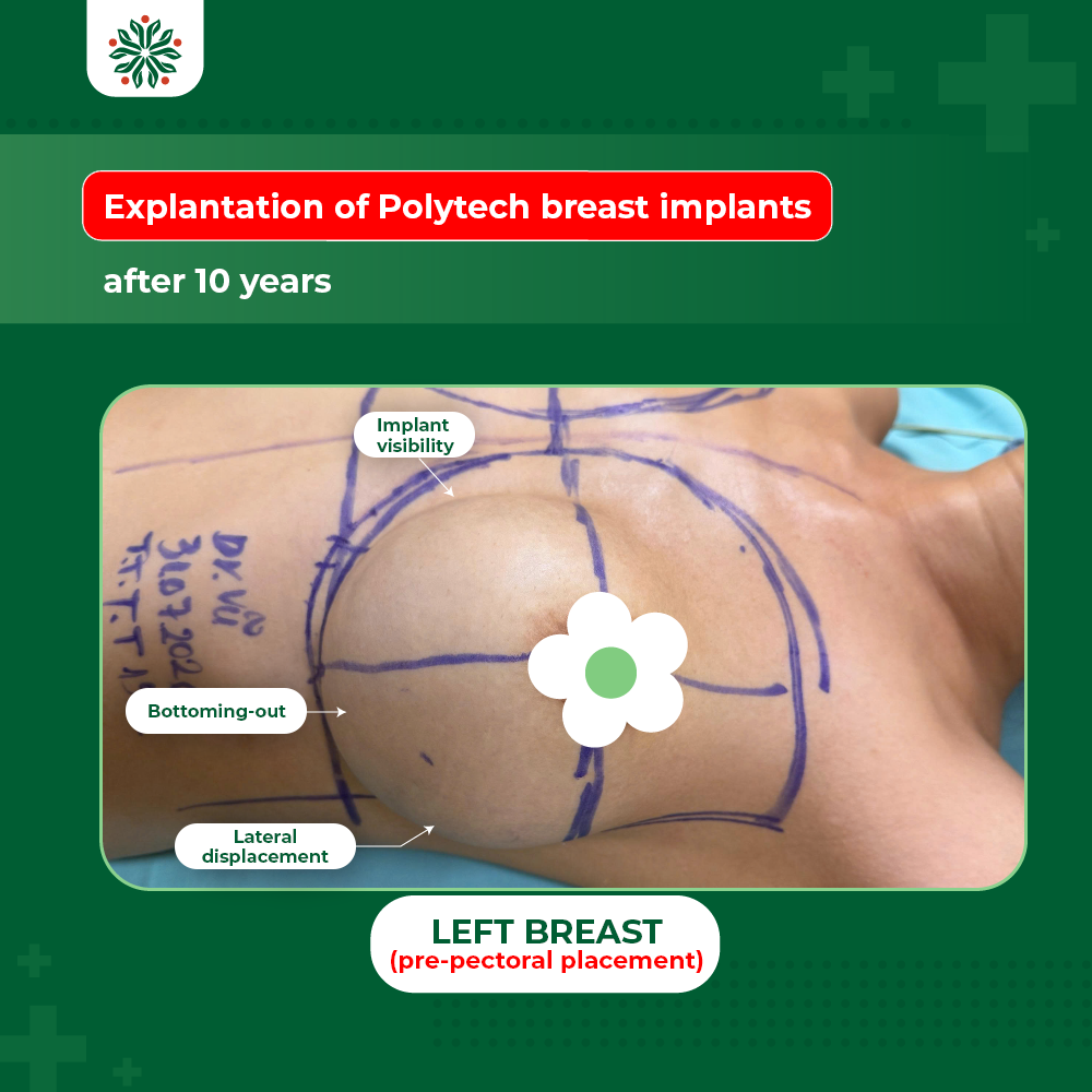
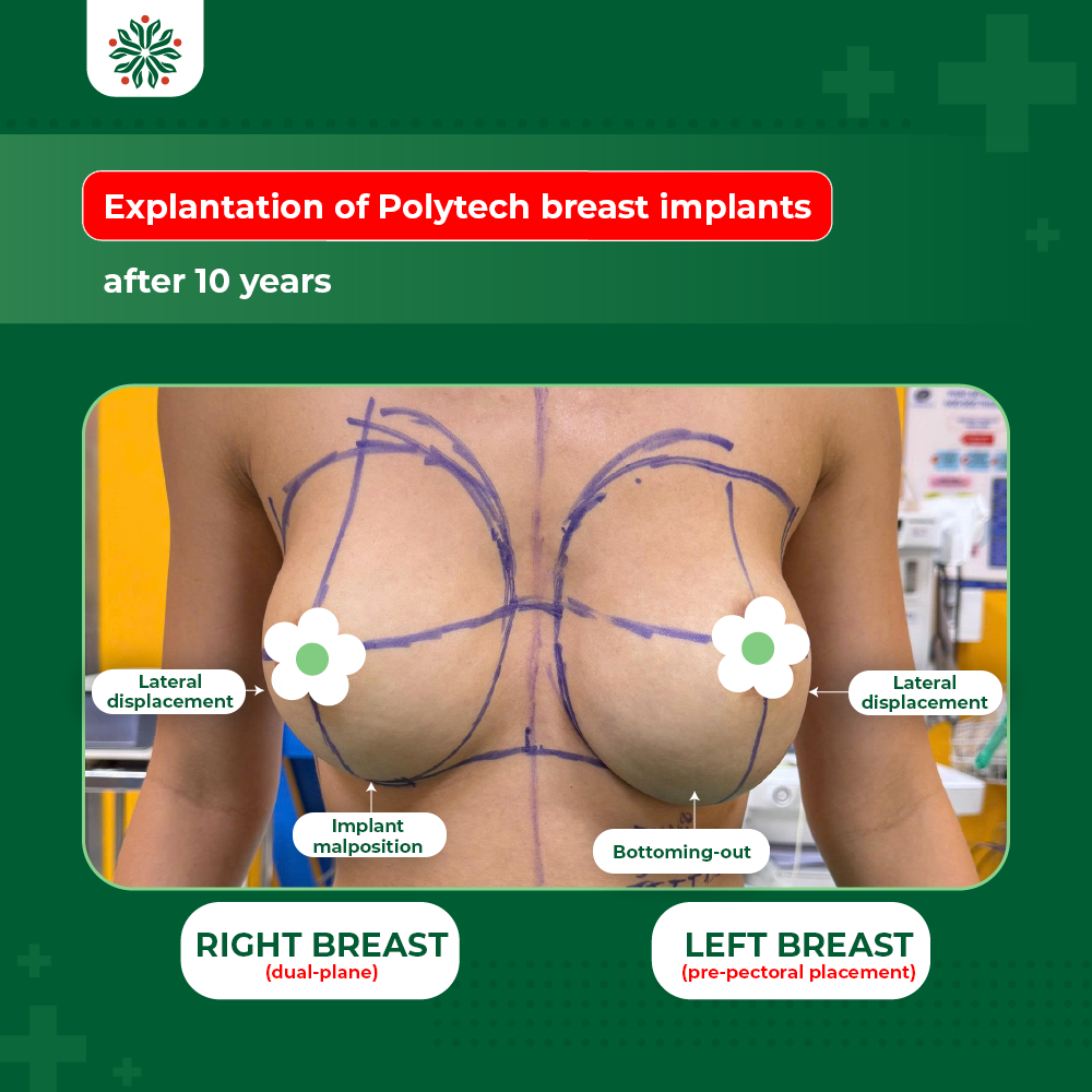
In patients with hypomastia, along with thin glandular and soft-tissue coverage, pre-pectoral placement—particularly in the subglandular plane—can increase the risk of visible implant edges, making the implant visually or palpably apparent through the skin. This is especially pronounced in women after childbirth, during lactation, or in a supine position. The primary reason is that the implant lacks coverage from the pectoralis major muscle and is solely covered by glandular tissue, adipose tissue, and skin. When this soft-tissue layer is excessively thin, irregular fibrosis reduces its coverage capacity, and the fibrotic glandular tissue exerts varying pressure across different areas of the implant. Consequently, the contour, edges, or folds of the implant become visible, compromising the natural appearance of the breast.
Challenges in Converting a Pre-Pectoral Pocket to a Sub-Pectoral Pocket
For cases with long-standing pre-pectoral placement (specifically subglandular and subfascial) presenting with thin coverage, stretched skin, implant malposition, bottoming-out, or malpositioned pockets, the primary challenge is the pre-existence of a pocket anterior to the pectoralis major muscle. If a new sub-pectoral pocket is created followed by immediate implant placement, the surgeon must meticulously manage the old pocket and the communication between the two spaces. Failure to control the old pocket increases the risk of the implant migrating back into the old pocket, resulting in malposition, a double-bubble deformity, or exacerbated breast contour distortion.
Additionally, the pectoralis major muscle provides only partial coverage of the implant. In cases of prolonged subglandular placement, the glandular tissue and skin can become stretched, thinned, or lose elasticity. Therefore, when planning to convert the implant from a subglandular to a sub-pectoral plane, the surgeon must thoroughly evaluate glandular tissue thickness, the degree of aging and elasticity of the glandular tissue and skin, the condition of the old pocket, as well as the size, diameter, and projection of the new implant.
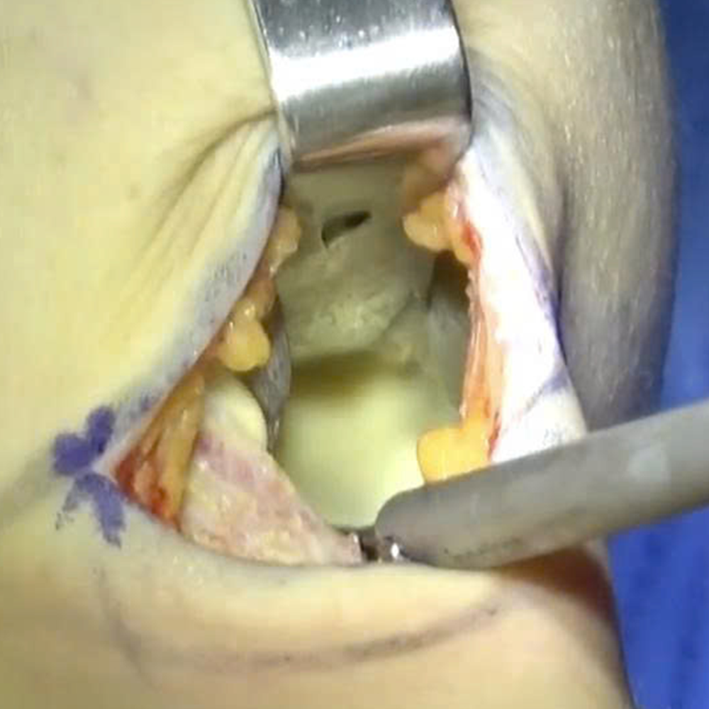
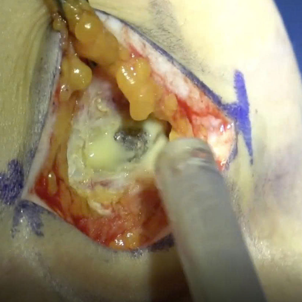
In cases involving significant aging of the glandular and periglandular tissues, excess skin, and accompanying complications such as implant malposition, bottoming-out, lateral displacement, and capsular contracture, the surgeon may opt for explantation first. Waiting approximately 6 months for the tissue and breast pocket to stabilize allows the surgeon to re-evaluate the breast structure and select an appropriate implant size and placement location prior to the subsequent surgery, thereby ensuring optimal aesthetic outcomes and minimizing complications. Furthermore, muscle fibrosis, which is noted in many pre-pectoral placement cases, is another crucial factor that must be evaluated when converting the pocket from anterior to posterior of the pectoralis major muscle in a single-stage procedure.
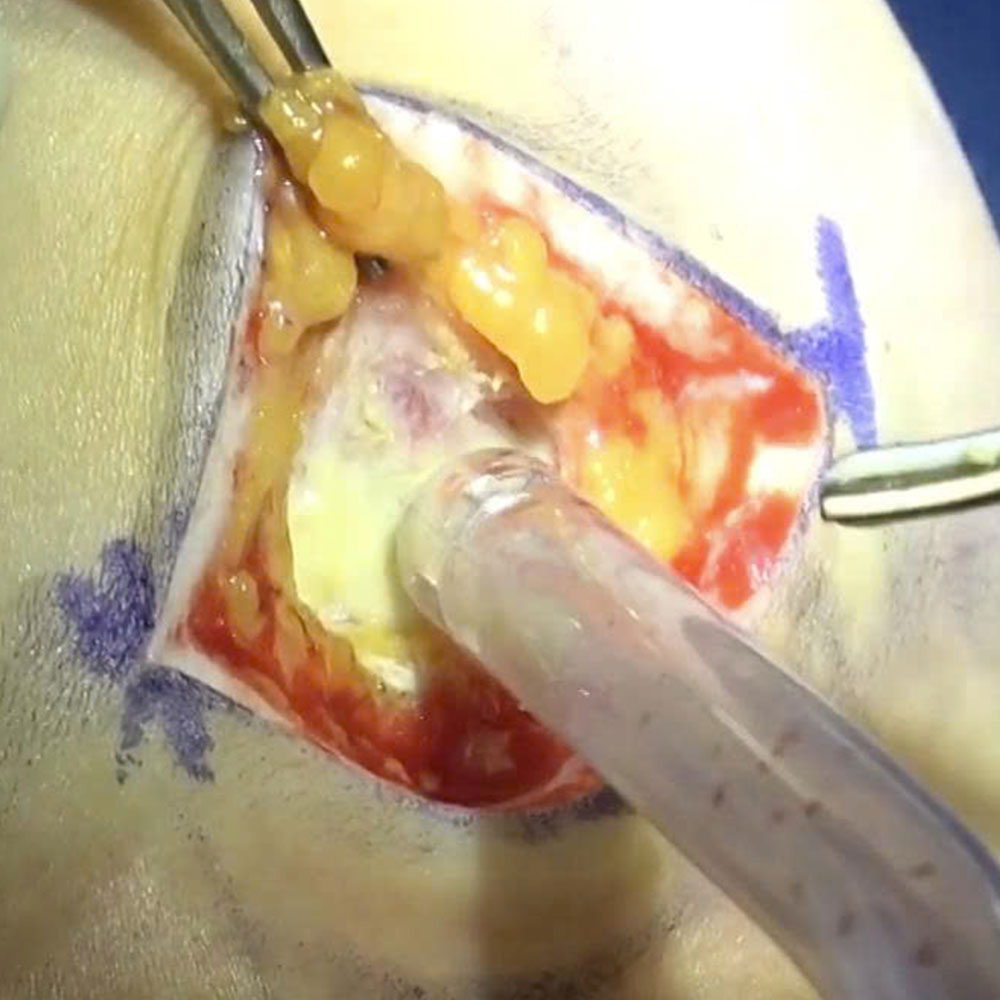
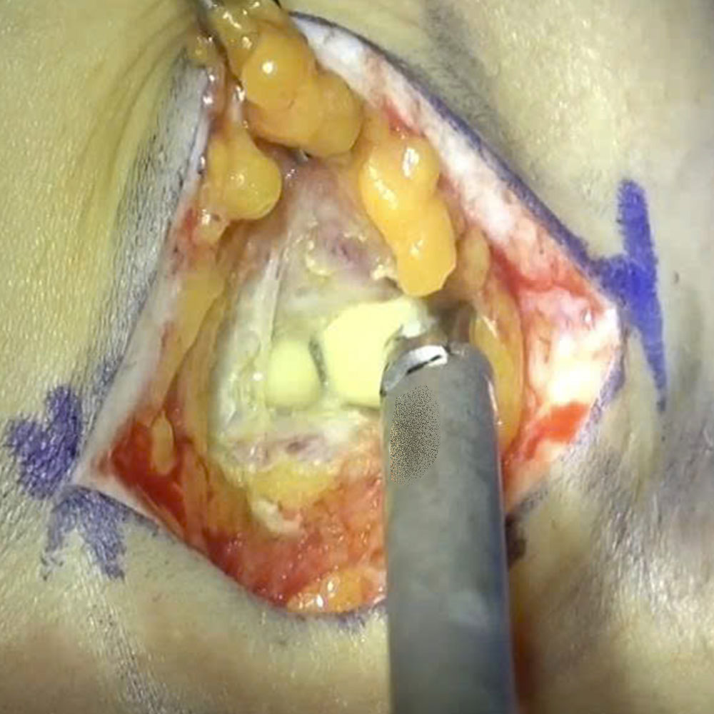
During the explantation of textured Polytech implants and pocket management using an ultrasonic scalpel, the surgeon identified different placement planes between the two breasts. On the right side, the intact 315 cc Polytech implant was placed in a dual-plane, with a portion of the implant situated under the pectoralis major muscle; the pocket shell was thin, and the pocket was stretched, exhibiting malposition and bottoming-out at the lateral border and inframammary fold (IMF). On the left side, the implant was placed in the subglandular plane, and the underlying pectoralis major muscle remained intact. Upon opening the pocket, the surgeon noted turbid yellow fluid and swollen, edematous, and bleeding-prone pocket tissue; the size specifications on the old implant were illegible, and the implant shell exhibited a dark yellow discoloration, indicating more advanced aging compared to the right implant.
These intraoperative findings demonstrate a marked discrepancy in the placement planes, pocket conditions, and periprosthetic tissues between the two breasts after years of augmentation. In the case of Ms. Hien, Dr. Vu elected against placing a new implant and performing an immediate pocket conversion. Instead, he chose the optimal approach of waiting 6 to 12 months for the tissue structures to stabilize. A sample of the turbid yellow fluid from the left breast pocket was collected for bacterial culture and susceptibility testing. The specimen was documented as purulent fluid from the left implant pocket.
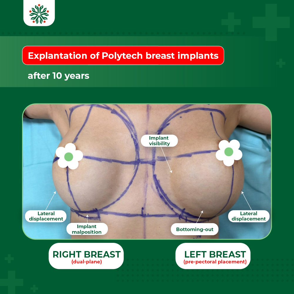
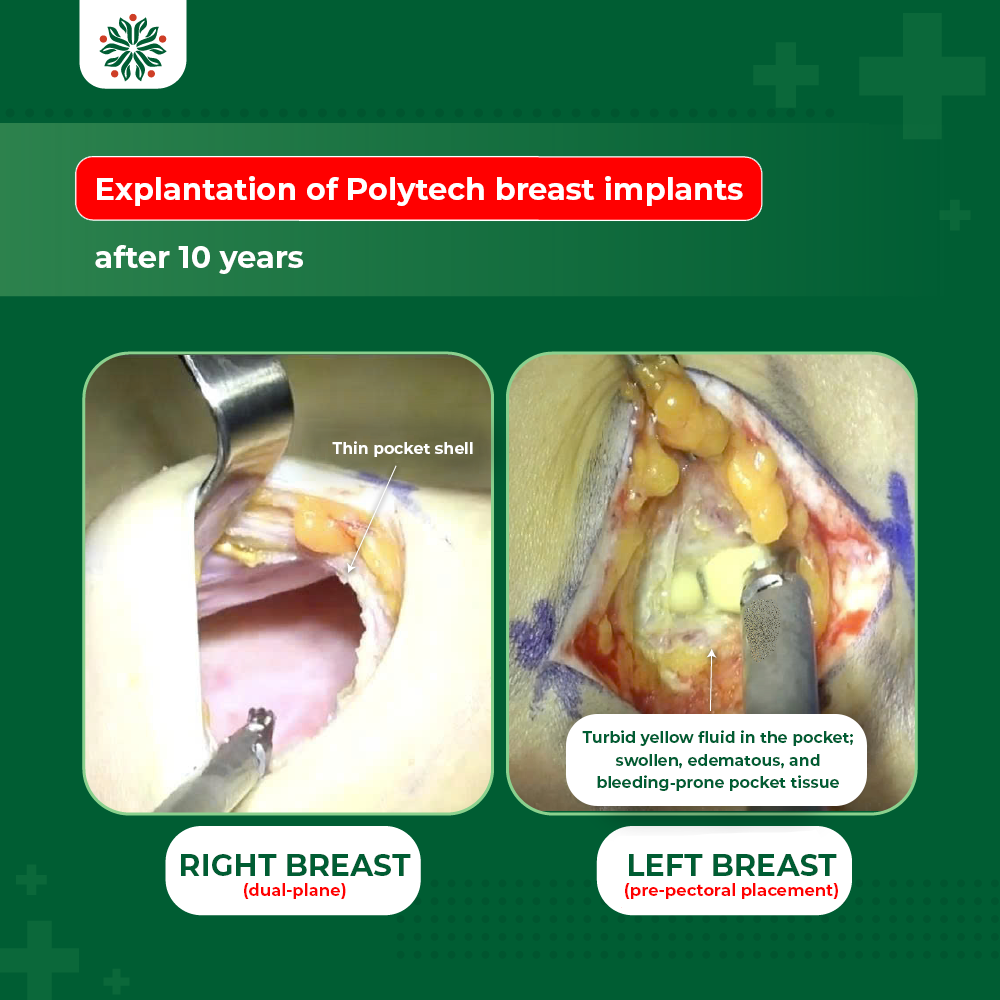
The FDA states that breast implants are not lifetime devices and the risk of complications increases over time. Therefore, to achieve long-term stability, the selection of the placement plane must be personalized based on thoracic anatomy, the thickness and fibrotic degree of the glandular tissue, pectoralis muscle characteristics, implant size, and the feasibility of creating an appropriate pocket. Source: https://www.fda.gov/medical-devices/breast-implants/things-consider-getting-breast-implants
Currently, MSc, MD. Ho Cao Vu prioritizes the sub-pectoral placement plane. According to the surgeon, the advantage of this technique is the additional muscle layer providing coverage, meaning anterior tissue aging has less impact on the implant. Simultaneously, it allows the surgeon to actively control and reconstruct the pocket based on the patient’s anatomical features and desired breast contour.
Intraoperatively, the surgeon utilizes specialized retractors combined with an ultrasonic scalpel to dissect, release, and reshape the implant pocket. This aims to achieve an appropriate pressure distribution around the implant while minimizing tissue and muscle trauma during manipulation.
According to MSc, MD. Ho Cao Vu, the precise creation of a sub-pectoral pocket is also significant for cases requiring future revisional surgery. When a patient wishes to increase or decrease implant volume, or alter the diameter, projection, or overall breast contour, the surgeon can re-evaluate the existing pocket and select a revisional strategy tailored to the breast structure at that time.


